Copyright
©2010 Baishideng Publishing Group Co.
World J Gastroenterol. Nov 21, 2010; 16(43): 5416-5423
Published online Nov 21, 2010. doi: 10.3748/wjg.v16.i43.5416
Published online Nov 21, 2010. doi: 10.3748/wjg.v16.i43.5416
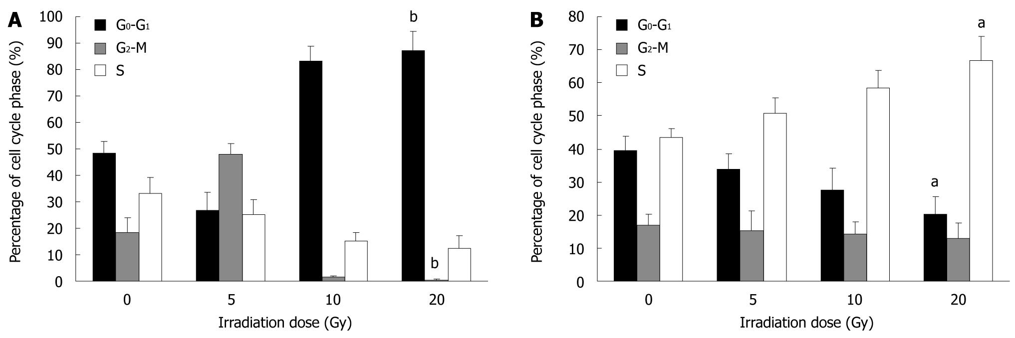
Figure 1 Flow cytometry of cell cycle response to irradiation in SW480 (A) and SW620 (B) cells.
aP < 0.05, bP < 0.01.
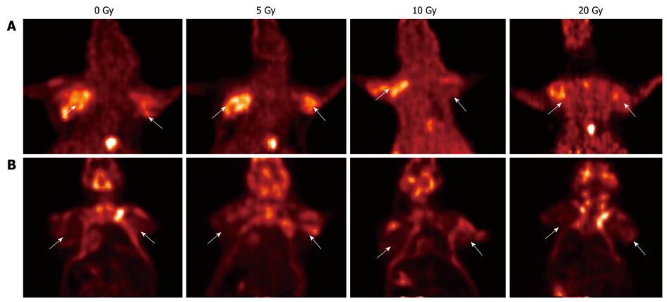
Figure 2 Coronal micro-position emission tomography sections of mice 60 min after injection with 18F-fluorothymidine (A) or 18F-fluorodeoxyglucose (B) before and 24 h after irradiation (3 mice/ group).
Implanted tumors (arrows) are located on the left (SW480) and the right (SW620) front legs of mice. Uptake of radiotracers was normalized to lung radiotracer uptake and expressed as ratio to non-tumor (T/NT) uptake.
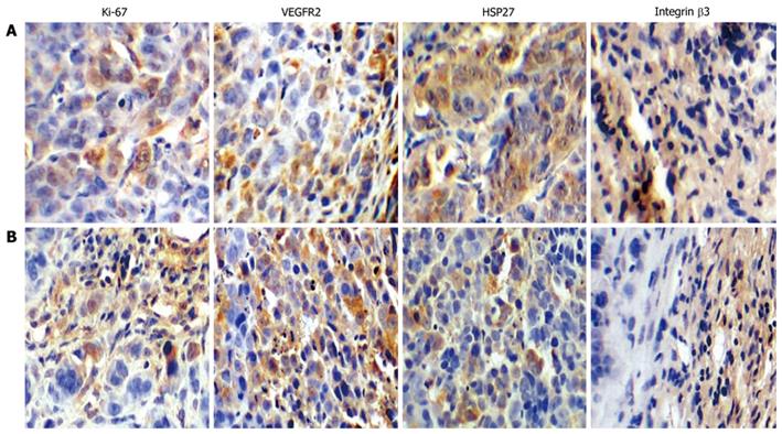
Figure 3 Immunohistochemical analysis of expression of integrin β3, K-i67, heat shock protein 27 and vascular endothelial growth factor receptor 2 protein in SW480 (A) and SW620 (B) cell lines by immunohistochemical detection (400 ×).
VEGFR2: Vascular endothelial growth factor receptor 2; HSP27: Heat shock protein 27.
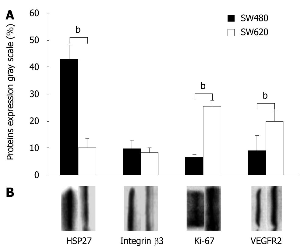
Figure 4 Western blotting analysis and quantitation of heat shock protein 27, integrin β3, Ki-67, and vascular endothelial growth factor receptor 2 protein expression in SW480 and SW620 tumors.
bP < 0.01. VEGFR2: Vascular endothelial growth factor receptor 2; HSP27: Heat shock protein 27.
- Citation: Wang H, Liu B, Tian JH, Xu BX, Guan ZW, Qu BL, Liu CB, Wang RM, Chen YM, Zhang JM. Monitoring early responses to irradiation with dual-tracer micro-PET in dual-tumor bearing mice. World J Gastroenterol 2010; 16(43): 5416-5423
- URL: https://www.wjgnet.com/1007-9327/full/v16/i43/5416.htm
- DOI: https://dx.doi.org/10.3748/wjg.v16.i43.5416


