Copyright
©2010 Baishideng Publishing Group Co.
World J Gastroenterol. Nov 14, 2010; 16(42): 5380-5387
Published online Nov 14, 2010. doi: 10.3748/wjg.v16.i42.5380
Published online Nov 14, 2010. doi: 10.3748/wjg.v16.i42.5380
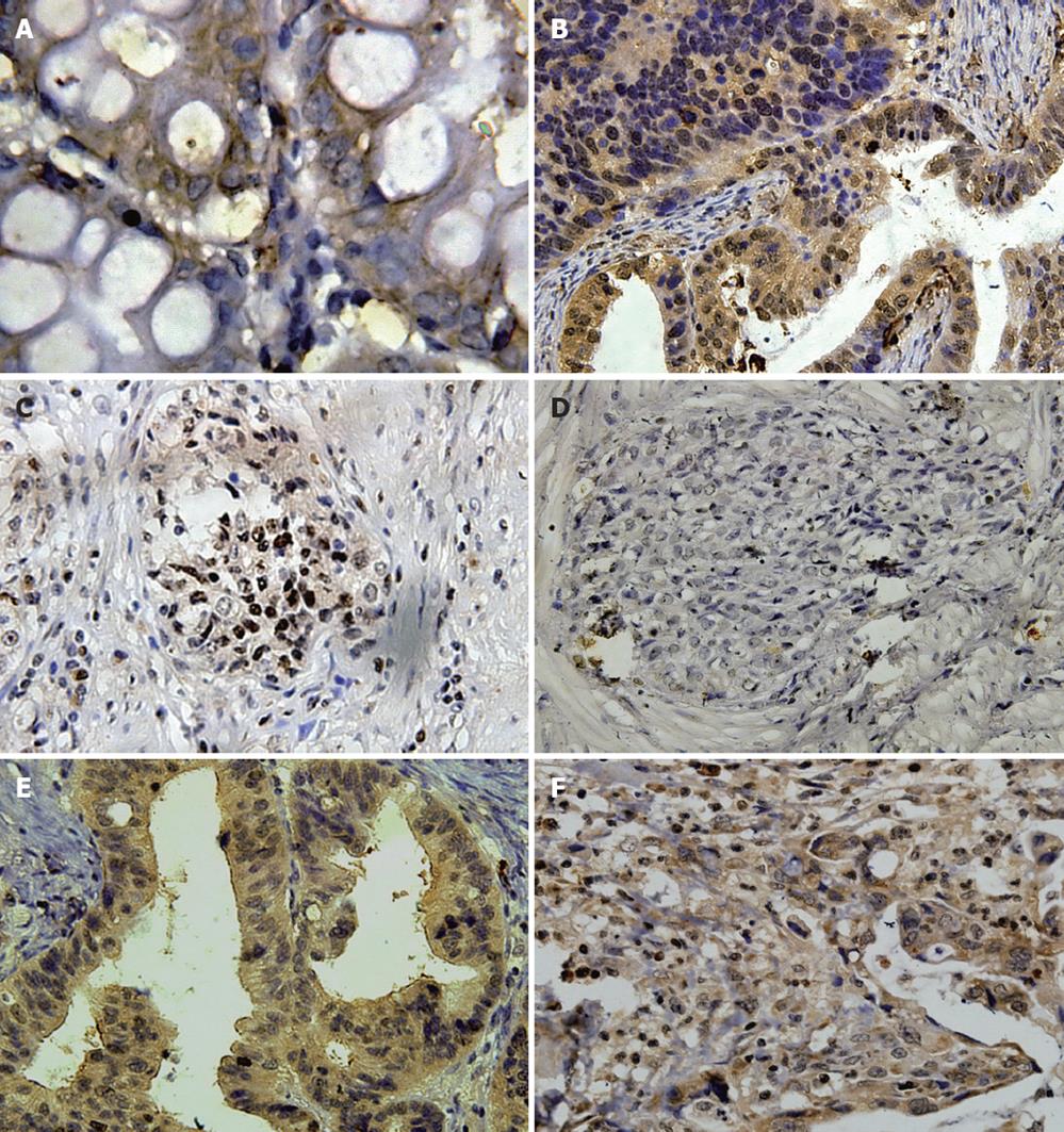
Figure 1 Expression in the cytoplasm of gastric cancer tissues (× 400).
A: Detection of signal transducer and activator of transcription-3 (STAT3) mRNA (in situ hybridization); B: STAT3 (IH); C: Phosphor-STAT3 (IH); D: Suppressor of cytokine signaling-1 (IH); E: Survivin (IH); F: Bcl-2 (IH).
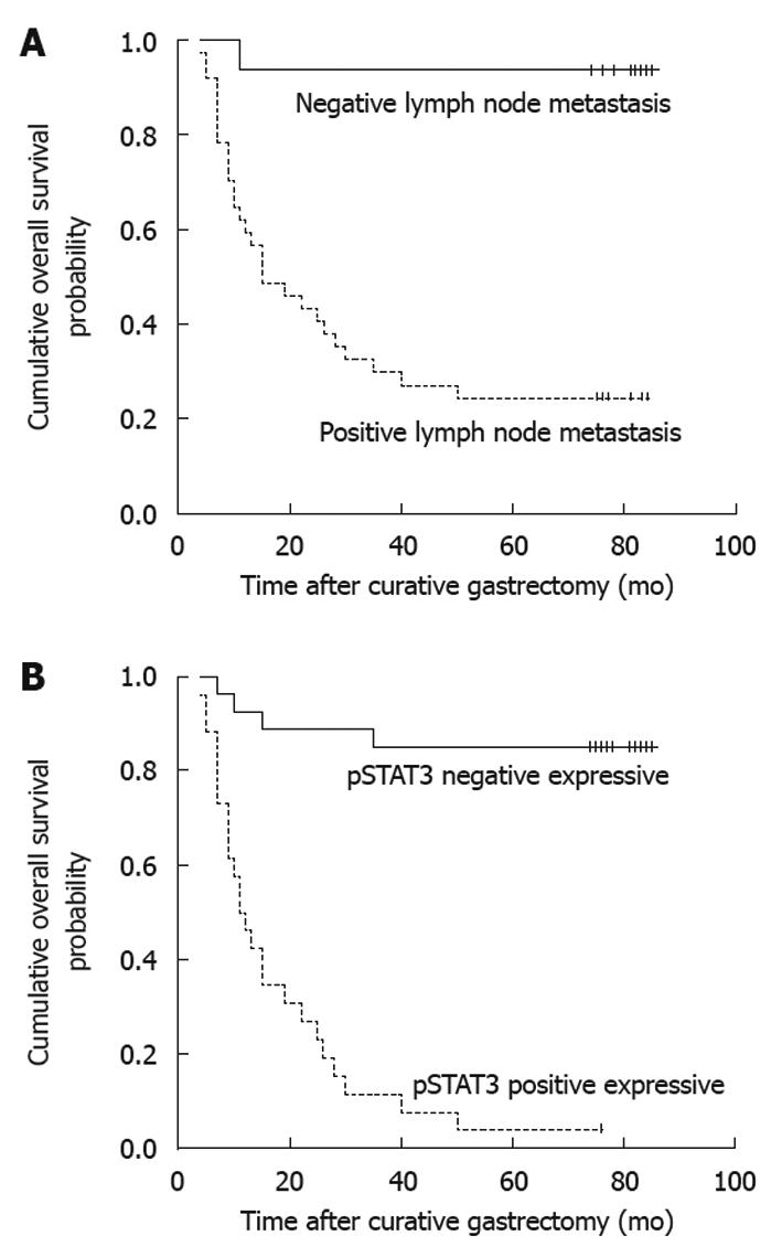
Figure 2 Survival curve in 53 gastric cancer patients following curative resection according to the metastatic status of lymph nodes (A) and different expression levels of phosphor-signal transducer and activator of transcription-3 (B).
A: Negative lymph node metastasis and positive lymph node metastasis; B: Negative and positive expression of pSTAT3.
- Citation: Deng JY, Sun D, Liu XY, Pan Y, Liang H. STAT-3 correlates with lymph node metastasis and cell survival in gastric cancer. World J Gastroenterol 2010; 16(42): 5380-5387
- URL: https://www.wjgnet.com/1007-9327/full/v16/i42/5380.htm
- DOI: https://dx.doi.org/10.3748/wjg.v16.i42.5380


