Copyright
©2010 Baishideng Publishing Group Co.
World J Gastroenterol. Oct 28, 2010; 16(40): 5135-5138
Published online Oct 28, 2010. doi: 10.3748/wjg.v16.i40.5135
Published online Oct 28, 2010. doi: 10.3748/wjg.v16.i40.5135
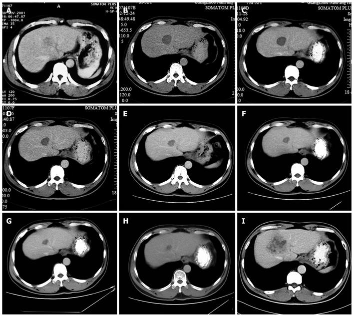
Figure 1 Computed tomography images of the hepatocellular carcinoma.
A: Tumor size was 3.5 cm × 3.2 cm before radiofrequency ablation (RFA) (2001-11); B: Tumor size was 3.2 cm × 3.1 cm at 12 mo after RFA (2002-12); C: Tumor size was 3.1 cm × 3.0 cm at 2 years after RFA (2003-11); D: Tumor size was 2.8 cm × 2.5 cm at 3 years after RFA (2005-1); E: Tumor size was 2.4 cm × 2.2 cm at 4 years after RFA (2005-11); F: Tumor size was 2.1 cm × 2.0 cm at 5 years after RFA (2006-09); G: Tumor size was 1.9 cm × 1.6 cm at 6 years after RFA (2007-11); H: Tumor size was 1.7 cm × 1.5 cm at 7 years after RFA (2008-11); I: Tumor size was 6.0 cm × 4.8 cm at 8 years after RFA (2009-12).
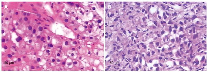
Figure 2 Pathological examinations (HE stain).
A: A well-differentiated hepatocellular carcinoma (HCC). Scale bar = 50 μm; B: A well-moderately differentiated HCC. Scale bar = 50 μm.
- Citation: Liao WJ, Shi M, Chen JZ, Li AM. Local recurrence of hepatocellular carcinoma after radiofrequency ablation. World J Gastroenterol 2010; 16(40): 5135-5138
- URL: https://www.wjgnet.com/1007-9327/full/v16/i40/5135.htm
- DOI: https://dx.doi.org/10.3748/wjg.v16.i40.5135


