Copyright
©2010 Baishideng Publishing Group Co.
World J Gastroenterol. Oct 28, 2010; 16(40): 5130-5134
Published online Oct 28, 2010. doi: 10.3748/wjg.v16.i40.5130
Published online Oct 28, 2010. doi: 10.3748/wjg.v16.i40.5130
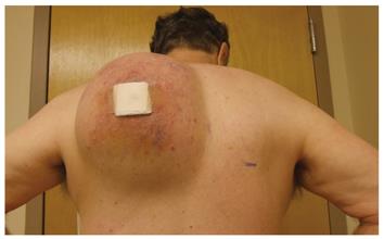
Figure 1 Large left shoulder mass.
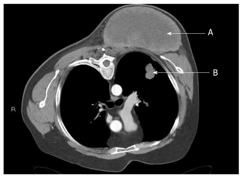
Figure 2 Chest computed tomography scan showing mass of left posterior chest (A) and suspected left pulmonary metastasis (B).
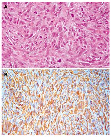
Figure 3 High-grade pleomorphic sarcoma show large pleomorphic tumor cells in a fibrous stroma with numerous mitotic figures (A), tumor cells show a strong diffuse cytoplasmic immunoreactivity to vimentin (B).
Other immunoreactions failed to discern any line of differentiation. The “vimentin only” immunophenotype leads to a diagnosis of undifferentiated pleomorphic sarcoma.
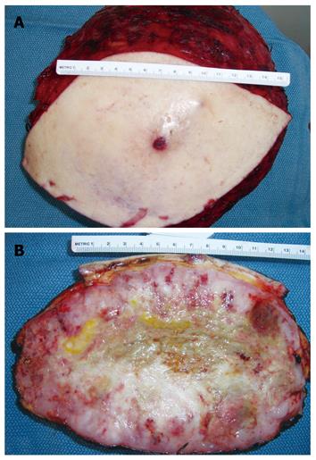
Figure 4 Resected left shoulder mass (A), cross section of resected shoulder mass showing central necrosis and smooth glistening dense fish flesh like tumor at the periphery (B).
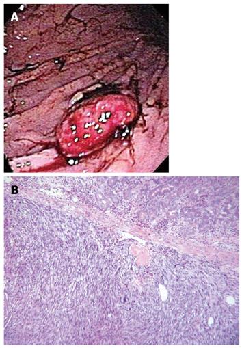
Figure 5 Endoscopic view of the stomach shows a mass with surface ulceration and blood clots (A), the photomicrograph (B) reveals a malignant neoplasm, composed of densely cellular stroma with spindle cells containing pleomorphic nuclei.
- Citation: Dent LL, Cardona CY, Buchholz MC, Peebles R, Scott JD, Beech DJ, Ballard BR. Soft tissue sarcoma with metastasis to the stomach: A case report. World J Gastroenterol 2010; 16(40): 5130-5134
- URL: https://www.wjgnet.com/1007-9327/full/v16/i40/5130.htm
- DOI: https://dx.doi.org/10.3748/wjg.v16.i40.5130


