Copyright
©2010 Baishideng Publishing Group Co.
World J Gastroenterol. Oct 14, 2010; 16(38): 4846-4853
Published online Oct 14, 2010. doi: 10.3748/wjg.v16.i38.4846
Published online Oct 14, 2010. doi: 10.3748/wjg.v16.i38.4846
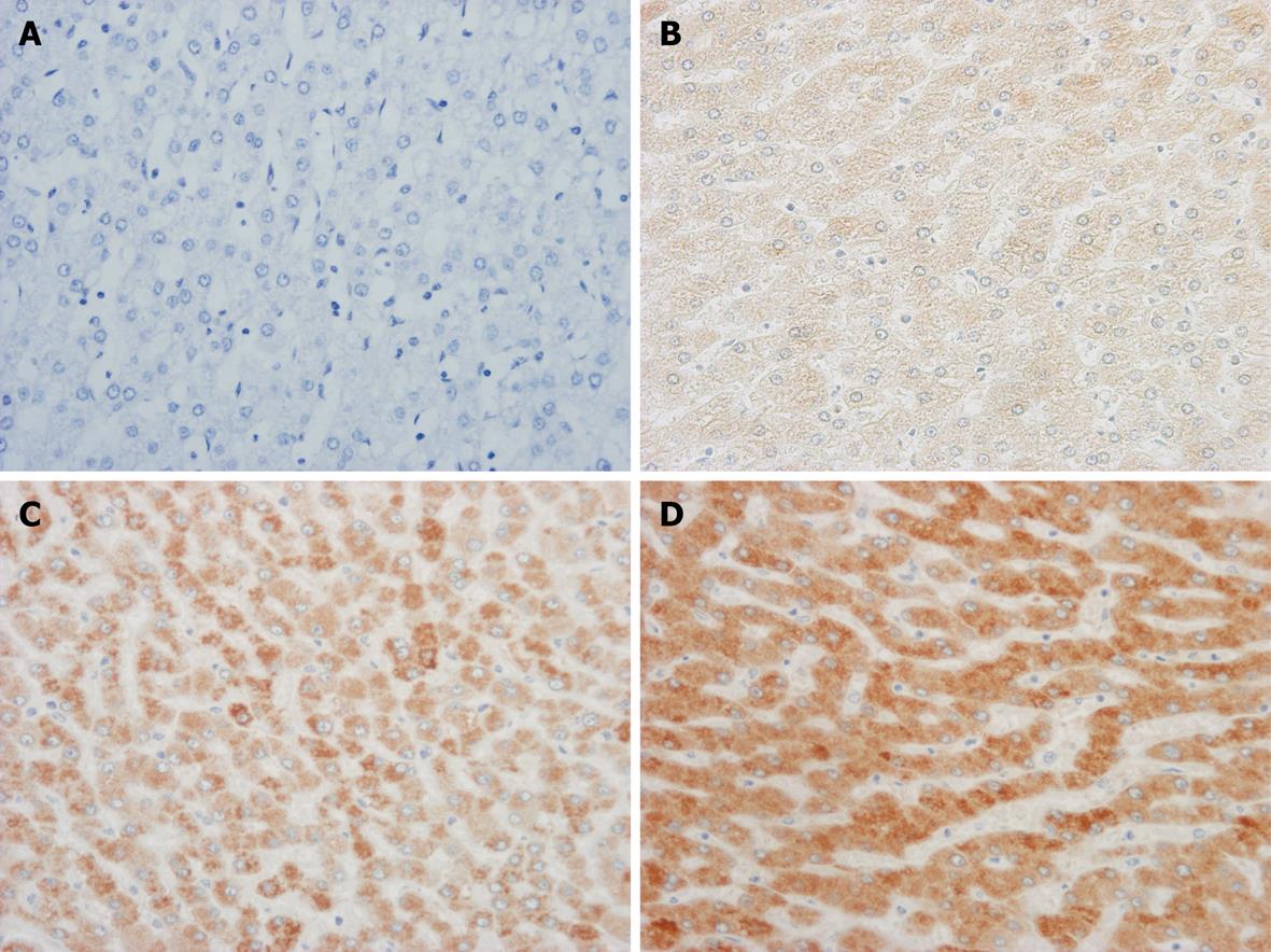
Figure 1 Grading of immunohistochemical staining for microsomal prostaglandin E synthase-1 protein in representative liver tissue (original magnification, × 200).
A: No immunoreactivity for microsomal prostaglandin E synthase-1 (mPGES-1) (grade 0); B: Weakly positive for mPGES-1 (grade 1); C: Moderately positive for mPGES-1 (grade 2); D: Strongly positive for mPGES-1 (grade 3).
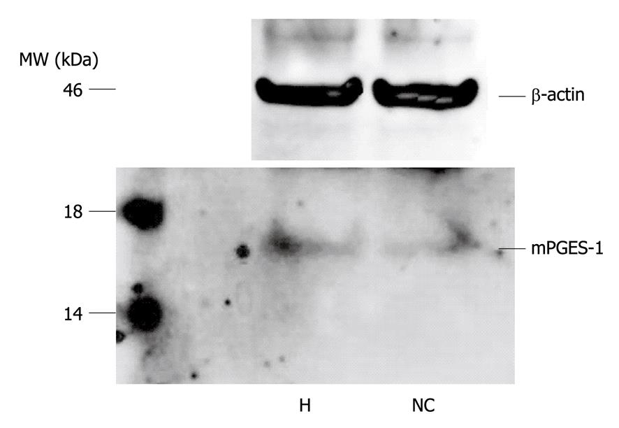
Figure 2 Western blotting analysis for microsomal prostaglandin E synthase-1.
A band of 16 kDa in molecular weight, thus indicating the presence of microsomal prostaglandin E synthase-1 (mPGES-1) protein, is identified in both hepatocellular carcinoma tumors (H) and non-cancerous liver (NC) tissues. MW: Molecular weight.
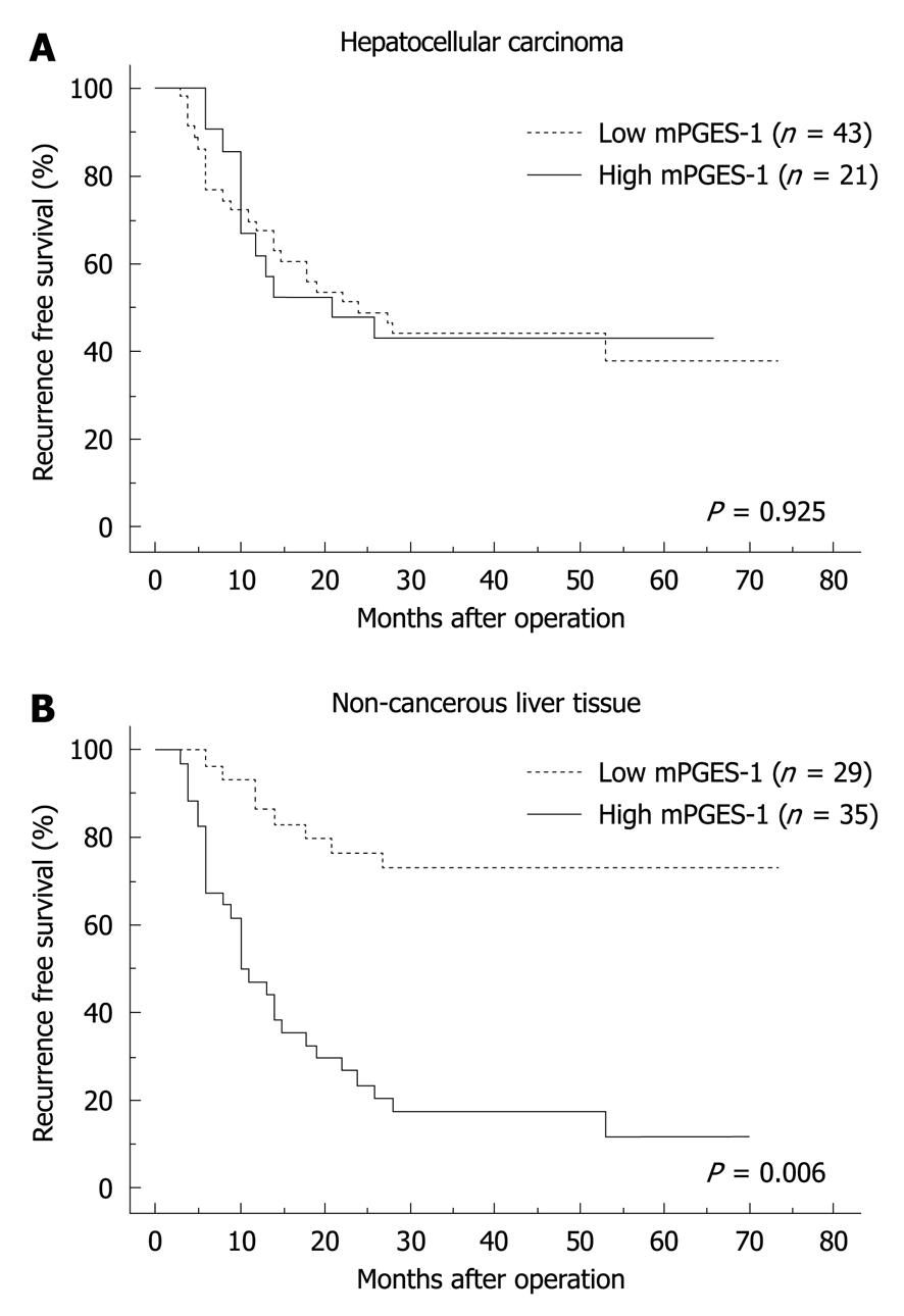
Figure 3 Recurrence-free survival time based on microsomal prostaglandin E synthase-1 expression in hepatocellular carcinoma tissues (A) and non-cancerous liver tissues (B).
The recurrence-free survival time is significantly shorter in patients with an increased expression of microsomal prostaglandin E synthase-1 (mPGES-1) in non-cancerous liver tissues.
- Citation: Nonaka K, Fujioka H, Takii Y, Abiru S, Migita K, Ito M, Kanematsu T, Ishibashi H. mPGES-1 expression in non-cancerous liver tissue impacts on postoperative recurrence of HCC. World J Gastroenterol 2010; 16(38): 4846-4853
- URL: https://www.wjgnet.com/1007-9327/full/v16/i38/4846.htm
- DOI: https://dx.doi.org/10.3748/wjg.v16.i38.4846


