Copyright
©2010 Baishideng Publishing Group Co.
World J Gastroenterol. Aug 28, 2010; 16(32): 4047-4054
Published online Aug 28, 2010. doi: 10.3748/wjg.v16.i32.4047
Published online Aug 28, 2010. doi: 10.3748/wjg.v16.i32.4047
Figure 1 Levels of ErbB2 expression in three cholangiocarcinoma cell lines and inhibition of ErbB2 phosphorylation by AG825.
A: Cells cultured in 10% fetal bovine serum (FBS)-containing medium were lysed and the lysates were analyzed for ErbB2 mRNA by real-time reverse-transcriptase polymerase chain reaction. Data are presented as mean ± SE of ErbB2 mRNA level normalized with β-actin mRNA obtained from three independent experiments; B: Cells were incubated without and with 30 μmol/L AG825 in 10% FBS medium for 6 h, then lysed and analyzed for ErbB2, phospho-ErbB2 (Y1248) and β-actin by Western blotting. The results shown are representative of three separate experiments. bP < 0.01 vs HuCCA-1 and KKU-100.
Figure 2 Invasion motility and proliferation of three cholangiocarcinoma cell lines.
A: Cells resuspended in 0.2% fetal bovine serum (FBS) medium were plated in the upper compartment of a Transwell chamber coated with and without Matrigel, for invasion and motility assays. The lower compartment was filled with 10% FBS medium. After 6 h incubation, numbers of invading/motile cells were counted; B: Cells were plated on 96-well plates in 10% FBS medium. After incubation for 12-72 h, cell survival was determined by 3-(4,5-dimethylthiazol-2-yl)-2,5-diphenyltetrazolium bromide assay. Data are presented as mean ± SE of the percentage relative to controls obtained from three separate experiments.
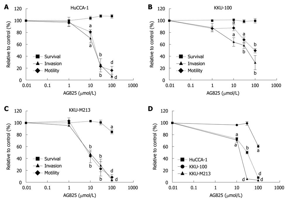
Figure 3 Effects of AG825 on cholangiocarcinoma cell invasion, motility and proliferation.
A-C: Cells resuspended in medium that contained 0.2% fetal bovine serum (FBS) and indicated concentrations of AG825 were plated in the upper compartment of a Transwell chamber coated with and without Matrigel, for invasion and motility assays. After 6 h incubation, numbers of invading/motile cells were counted. Cells treated with the same vehicle were used as controls. Cell survival after 6 h of AG825 treatment was determined by 3-(4,5-dimethylthiazol-2-yl)-2,5-diphenyltetrazolium bromide (MTT) assay; D: Cells were treated with various concentrations of AG825 in 10% FBS medium for 72 h, and cell survival was determined using the MTT assay. Data are presented as mean ± SE of the percentage relative to the controls obtained from three separate experiments, each done in duplicate. aP < 0.05, bP < 0.01, dP < 0.001 vs control.
Figure 4 Inhibition of ErbB2 expression by siRNA against ErbB2.
A: KKU-M213 cells were transfected with siErbB2 and the levels of ErbB2 at 0-96 h after transfection were determined by Western blotting; B: After 72 h of siRNA transfection, ErbB2 expression in HuCCA-1, KKU-100, and KKU-M213 cells was determined. Scramble siRNA (siNeg) was used as a negative control. The results shown are representatives of two (A) and three (B) separate experiments.
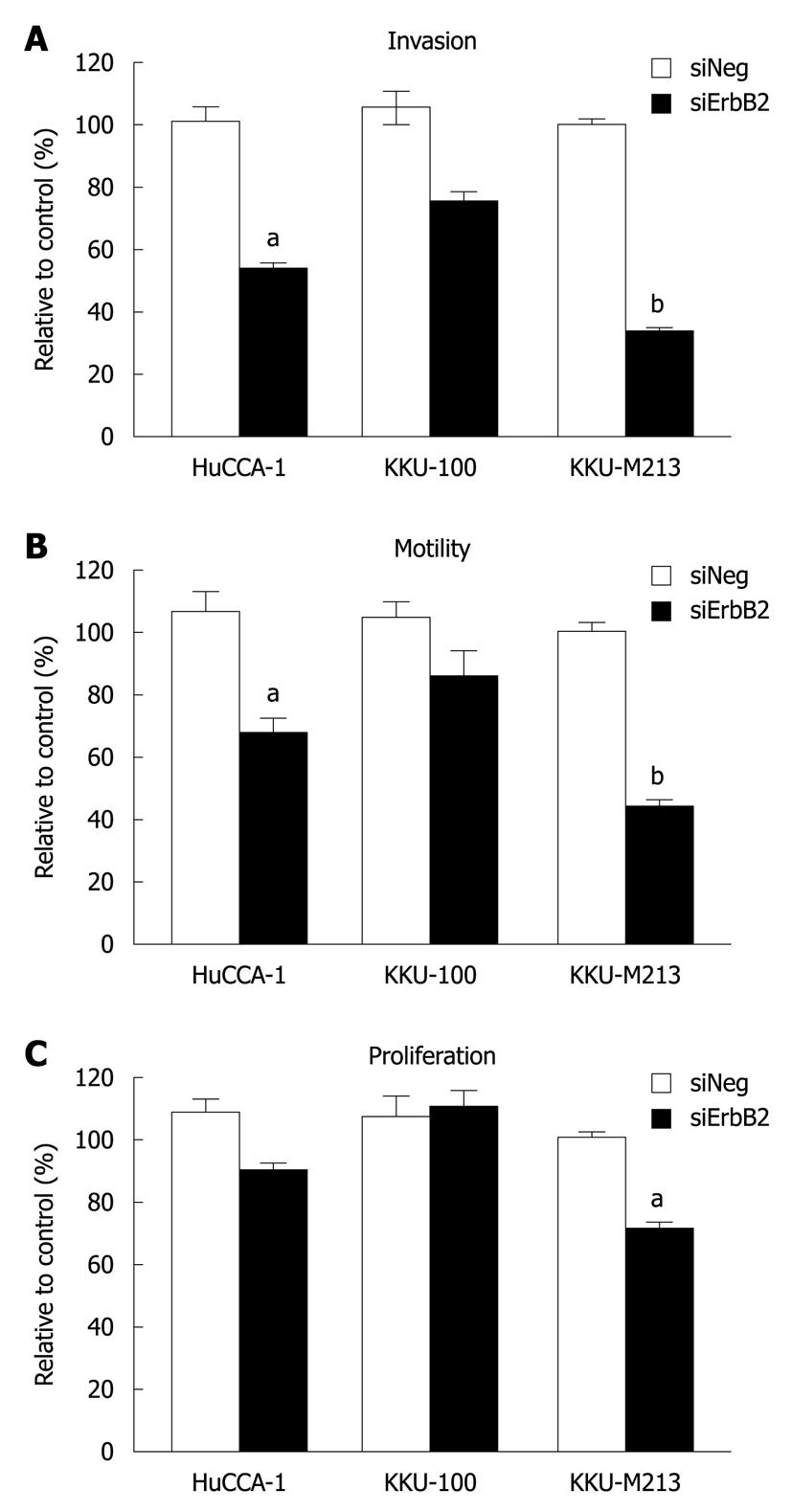
Figure 5 Effects of ErbB2 knockdown on cell invasion motility and proliferation of the three cholangiocarcinoma cell lines.
Cells transfected with siRNA (negative control and ErbB2 targeted siRNA) for 72 h were analyzed for in vitro invasion (A) and motility (B) by using a Transwell chamber coated with and without Matrigel. After 6 h of incubation, numbers of invading/motile cells were counted. Cell proliferation (C) was analyzed by determining the number of viable cells during 24-96 h post-transfection by 3-(4,5-dimethylthiazol-2-yl)-2,5-diphenyltetrazolium bromide assay. Data are expressed as mean ± SE of the percentage relative to the controls obtained from three independent experiments. aP < 0.05, bP < 0.01 vs controls.
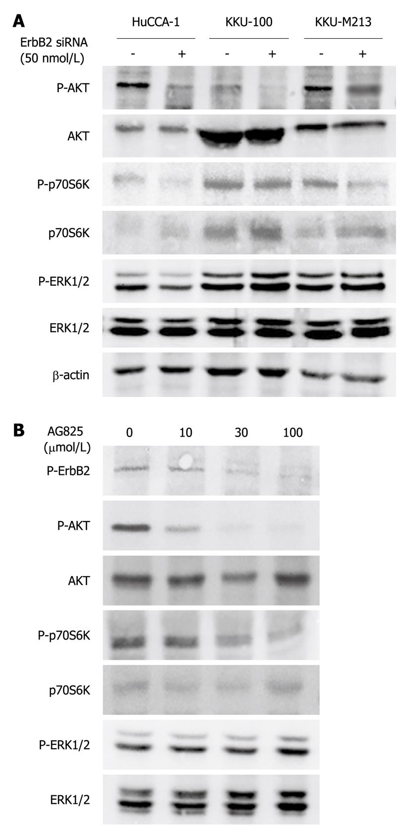
Figure 6 Effects of ErbB2 inhibition (using siRNA and specific kinase inhibitor) on ERK1/2, AKT, and p70S6K phosphorylation in the cholangiocarcinoma cell lines.
A: Cholangiocarcinoma cell lines were transfected with siErbB2 and siNeg, as a control, for 72 h and the collected cell lysates were analyzed by Western blotting; B: KKU-M213 cells were treated without and with AG825 in medium that contained 10% fetal bovine serum for 6 h, then lysed and analyzed by Western blotting. The results shown are representatives of three separate experiments.
- Citation: Treekitkarnmongkol W, Suthiphongchai T. High expression of ErbB2 contributes to cholangiocarcinoma cell invasion and proliferation through AKT/p70S6K. World J Gastroenterol 2010; 16(32): 4047-4054
- URL: https://www.wjgnet.com/1007-9327/full/v16/i32/4047.htm
- DOI: https://dx.doi.org/10.3748/wjg.v16.i32.4047


