Copyright
©2010 Baishideng Publishing Group Co.
World J Gastroenterol. Aug 28, 2010; 16(32): 4031-4038
Published online Aug 28, 2010. doi: 10.3748/wjg.v16.i32.4031
Published online Aug 28, 2010. doi: 10.3748/wjg.v16.i32.4031
Figure 1 Scanning electron microscopy pictures of the stratum compactum surface (A) and the abluminal surface (B) of small-intestinal submucosa.
Magnification × 500.
Figure 2 Scanning electron microscopy pictures of the surface structure of the samples with surface characteristics of the different original biomaterials.
A: Porcine dermal matrix; B: Porcine pericardial matrix; C: Bovine pericardial matrix. Magnification × 500.
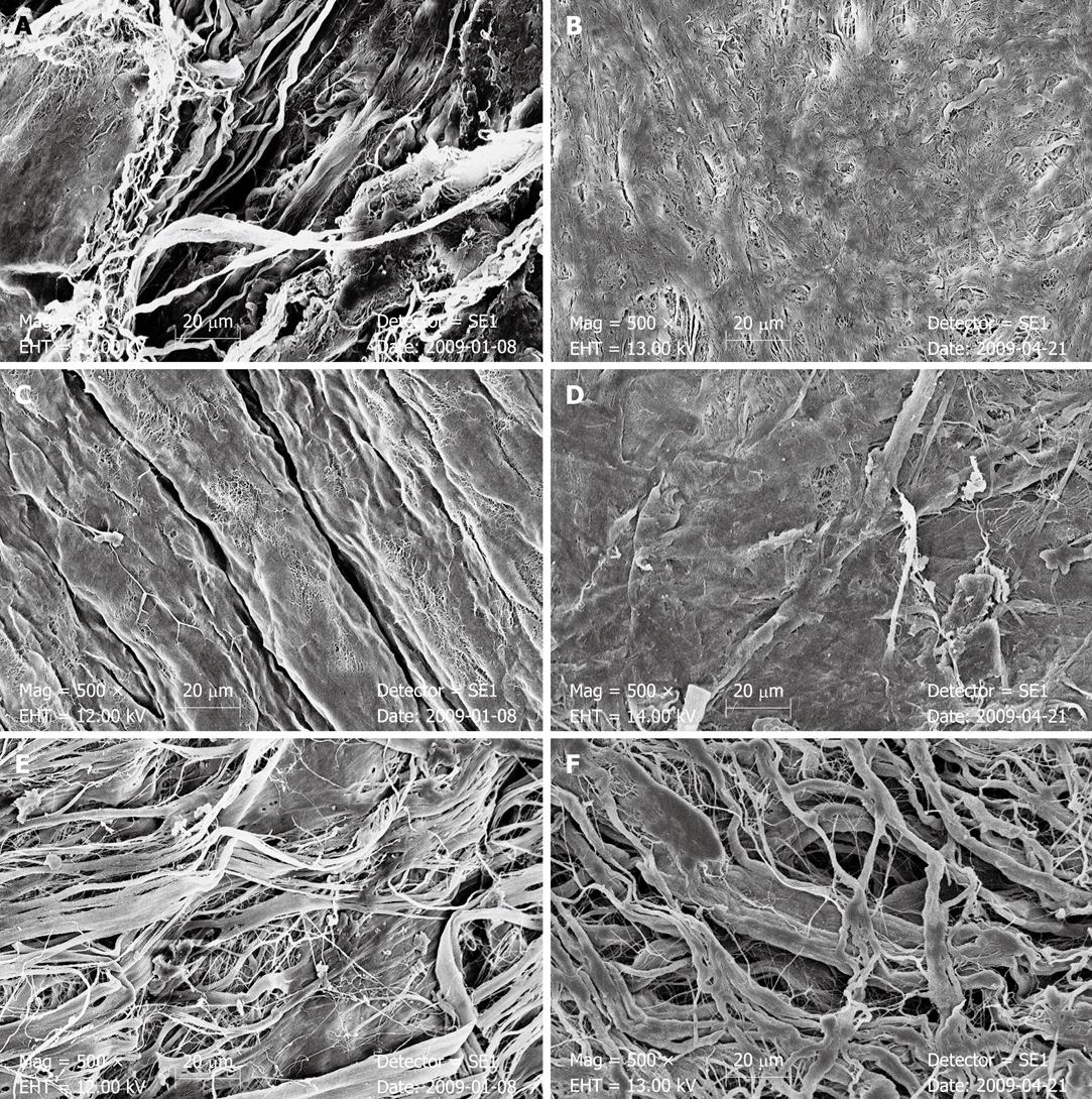
Figure 3 Scanning electron microscopy pictures of small-intestinal submucosa, porcine pericardial matrix and bovine pericardial matrix after 7 d of bile or pancreatic juice incubation.
A: Small-intestinal submucosa (SIS) incubated in bile; B: SIS incubated in pancreatic juice; C: Porcine pericardial matrix (PPM) incubated in bile; D: PPM incubated in pancreatic juice; E: Bovine pericardial matrix (BPM) incubated in bile; F: BPM incubated in pancreatic juice.
-
Citation: Hoeppner J, Marjanovic G, Helwig P, Hopt UT, Keck T. Extracellular matrices for gastrointestinal surgery:
Ex vivo testing and current applications. World J Gastroenterol 2010; 16(32): 4031-4038 - URL: https://www.wjgnet.com/1007-9327/full/v16/i32/4031.htm
- DOI: https://dx.doi.org/10.3748/wjg.v16.i32.4031


