Copyright
©2010 Baishideng.
World J Gastroenterol. Jan 21, 2010; 16(3): 398-402
Published online Jan 21, 2010. doi: 10.3748/wjg.v16.i3.398
Published online Jan 21, 2010. doi: 10.3748/wjg.v16.i3.398
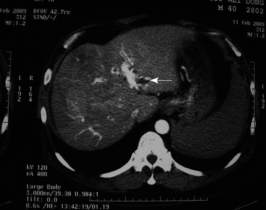
Figure 1 Computed tomography (CT) scan obtained before the first Transcatheter arterial chemoembolization (TACE) procedure.
Portal vein was shown during the arterial period, which suggested that the tumor embolus invaded the portal vein (arrow).
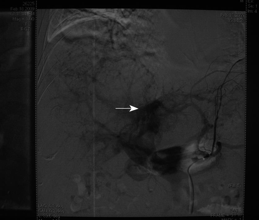
Figure 2 First TACE procedure.
Arterial-portal fistula (arrow).
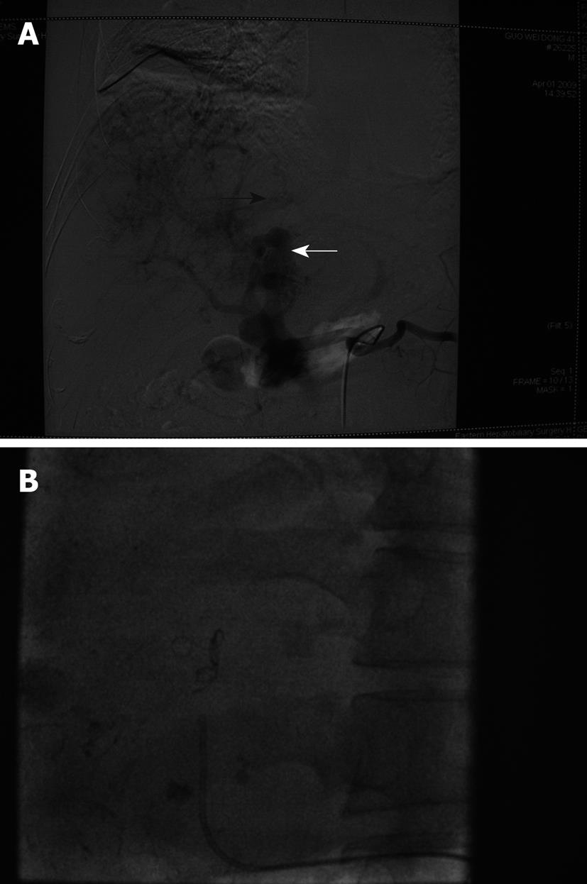
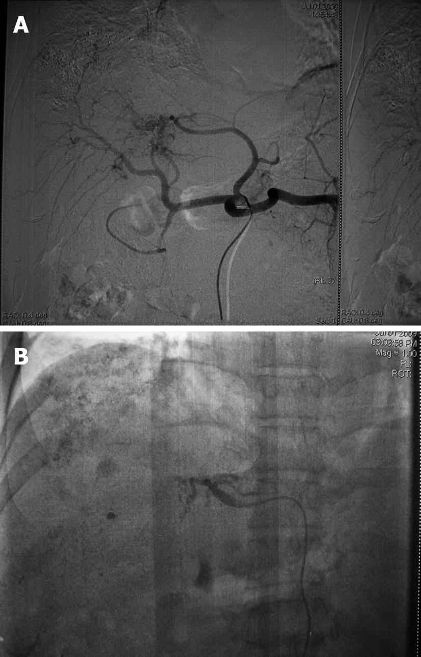
Figure 3 Second TACE procedure.
A: Hepatic arteriovenous fistula (black arrow), arterial-portal fistula (white arrow); B: Coils (5-8 mm).
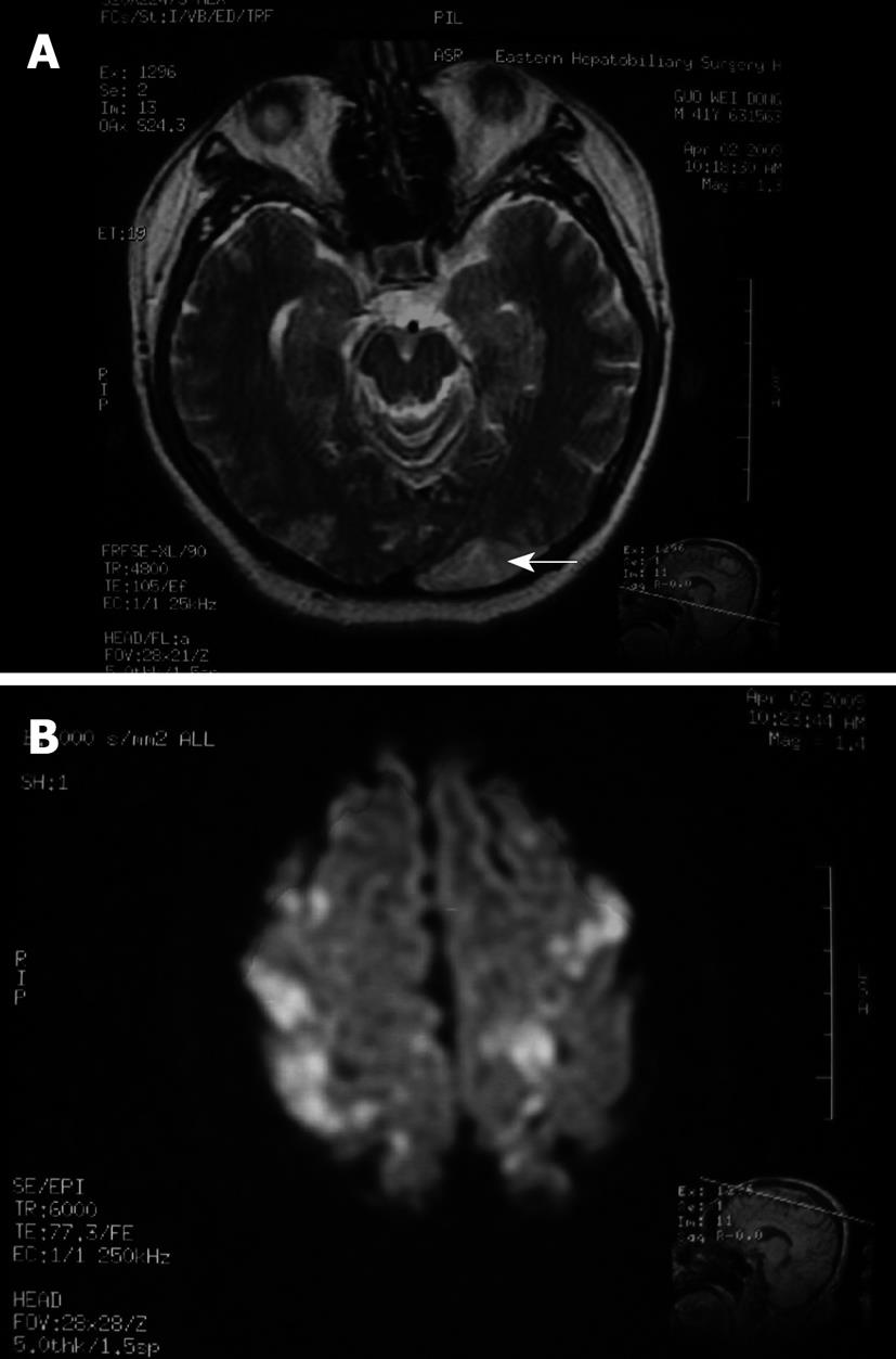
Figure 4 Cranial magnetic resonance imaging (MRI) obtained after the second TACE procedure.
A: cerebral lipiodol embolism (arrow); B: cerebral lipiodol embolism (multiple high signals).
Figure 5 Third TACE procedure.
A: Hepatic arteriovenous fistula was not seen, and the arterial-portal fistula was still present but improved; B: Lipiodol and gelatin sponge were injected through the left gastric artery and the arterial-portal fistula was embolized.
- Citation: Wu L, Yang YF, Liang J, Shen SQ, Ge NJ, Wu MC. Cerebral lipiodol embolism following transcatheter arterial chemoembolization for hepatocellular carcinoma. World J Gastroenterol 2010; 16(3): 398-402
- URL: https://www.wjgnet.com/1007-9327/full/v16/i3/398.htm
- DOI: https://dx.doi.org/10.3748/wjg.v16.i3.398


