Copyright
©2010 Baishideng.
World J Gastroenterol. Aug 7, 2010; 16(29): 3723-3726
Published online Aug 7, 2010. doi: 10.3748/wjg.v16.i29.3723
Published online Aug 7, 2010. doi: 10.3748/wjg.v16.i29.3723
Figure 1 Intraoperative cholangiography.
A: The right anterior segmental duct (RASD) emptying into the cystic duct is seen (arrow). The catheter is in the RASD. The right posterior segmental duct joining the left hepatic duct is also shown (arrowhead); B: After the left lateral segment was recovered, no stenosis was seen at the site of the repaired cystic duct (arrow). Cholangiography was performed through the stump of the left hepatic duct.
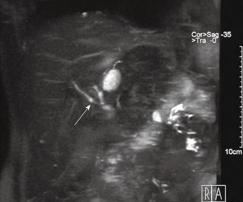
Figure 2 Magnetic resonance cholangiography was performed 3 mo postoperatively.
The right anterior segmental duct and cystic duct are clearly seen (arrow).
- Citation: Ishiguro Y, Hyodo M, Fujiwara T, Sakuma Y, Hojo N, Mizuta K, Kawarasaki H, Lefor AT, Yasuda Y. Right anterior segmental hepatic duct emptying directly into the cystic duct in a living donor. World J Gastroenterol 2010; 16(29): 3723-3726
- URL: https://www.wjgnet.com/1007-9327/full/v16/i29/3723.htm
- DOI: https://dx.doi.org/10.3748/wjg.v16.i29.3723


