Copyright
©2010 Baishideng.
World J Gastroenterol. Jul 28, 2010; 16(28): 3584-3591
Published online Jul 28, 2010. doi: 10.3748/wjg.v16.i28.3584
Published online Jul 28, 2010. doi: 10.3748/wjg.v16.i28.3584
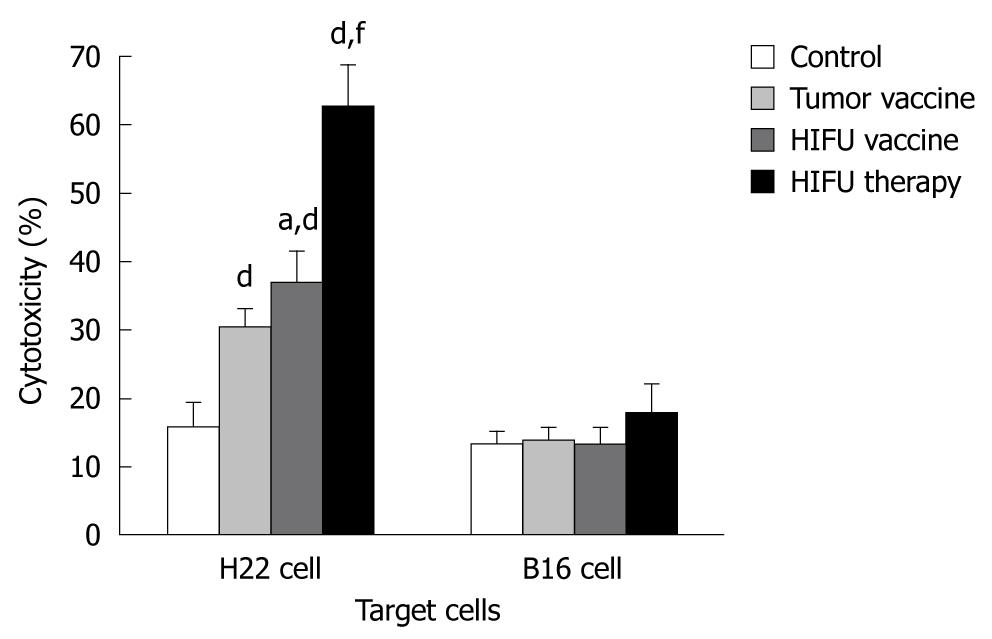
Figure 1 Cytotoxic activity of cytotoxic T lymphocytes against either H22 or B16 tumor cells in vitro at 10:1 effector:target ratio in the vaccinated and high-intensity focused ultrasound-treated mice.
Naïve mice were vaccinated with high-intensity focused ultrasound (HIFU)-generated and tumor-generated vaccines, and saline alone once a week for 2 wk. The mice bearing H22 tumors were also treated with HIFU ablation. The vaccinated animals were sacrificed 7 d after the 2nd vaccination, and the HIFU-treated mice were sacrificed 15 d after HIFU therapy. The spleens were harvested, and single cell suspensions were generated. The splenic lymphocytes were then co-cultured with either H22 or B16 cells for 24 h. The cytotoxicity of the cytotoxic T lymphocytes was determined with a 3-(4,5-dimethylthiazol-2-yl)-2,5-diphenyltetrazolium bromide assay in each group after 2 h co-incubation. aP < 0.05 vs the tumor-generated vaccine; dP < 0.001 vs the control; fP < 0.001 vs the HIFU- and tumor-generated vaccines.
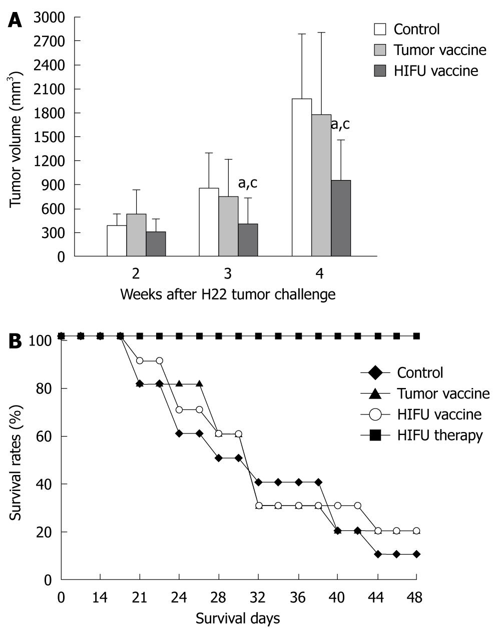
Figure 2 High-intensity focused ultrasound-generated vaccine inhibits tumor growth after a subsequent tumor challenge in a mouse H22 tumor model.
Naïve mice were vaccinated with high-intensity focused ultrasound (HIFU)-generated and tumor-generated vaccines, and saline alone once a week for 2 wk. The mice bearing H22 tumors were also treated with HIFU ablation. 7 d after the 2nd vaccination, the vaccinated animals were challenged with 2 × 10⁶ viable H22 cells, and the HIFU-treated mice received a second tumor challenge with the same number of H22 cells 15 d after HIFU therapy. Tumor diameters were measured for 3 wk, and the results were reported as the tumor volume. All mice were followed up for 48 d, and a cumulative survival rate was calculated in each group. A: The tumor volume, measured with a Vernier caliper, in the vaccinated and HIFU-treated mice after a subsequent tumor challenge. aP < 0.05 vs the control; cP < 0.05 vs the tumor-generated vaccine; B: Cumulative survival curves, calculated with the Kaplan-Meier method, in the vaccinated and HIFU-treated mice. Compared to the other groups, HIFU therapy shows a significant increase in survival (P < 0.001, the log-rank test).
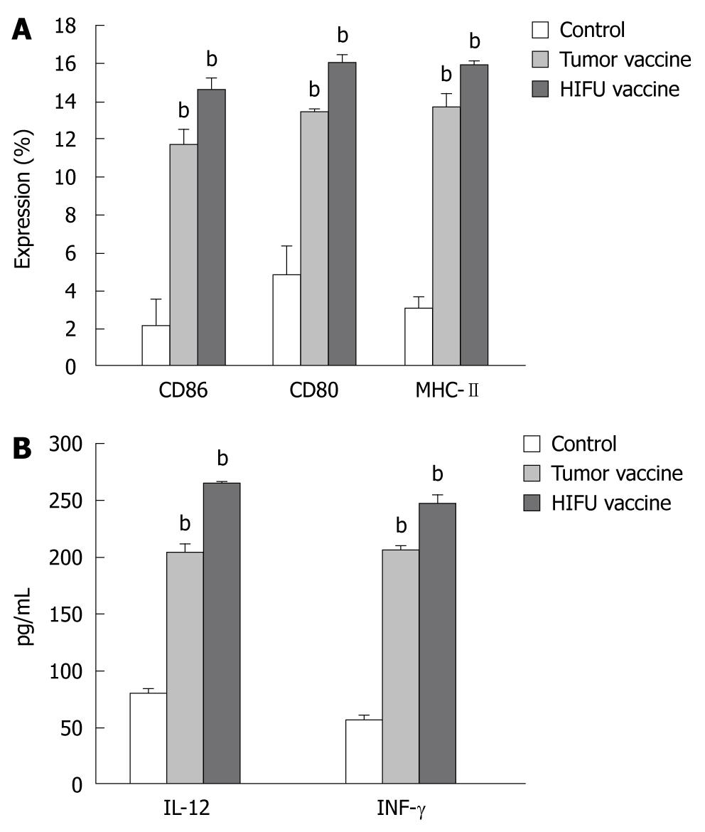
Figure 3 High-intensity focused ultrasound-generated and tumor-generated vaccines activate dendritic cells.
Immature dendritic cells (DCs) were isolated from C57BL/6J bone marrow cultures, and then incubated for 5 d with the high-intensity focused ultrasound (HIFU)-generated vaccine, tumor-generated vaccine, and mouse serum alone. After incubation the cells were subjected to flow cytometry. Culture supernatants were harvested, and the enzyme-linked immunosorbent assay method was used to determine the production of interleukin (IL)-12 and interferon (IFN)-γ in the supernatants. bP < 0.001 vs the control. A: HIFU-generated vaccine induces the maturation of bone marrow-derived DCs. Results are reported as percentage of MHC-II+, CD80+ and CD86+ cells in the total population; B: HIFU-generated vaccine induces IL-12 and IFN-γ secretion by mature DCs.
- Citation: Zhang Y, Deng J, Feng J, Wu F. Enhancement of antitumor vaccine in ablated hepatocellular carcinoma by high-intensity focused ultrasound. World J Gastroenterol 2010; 16(28): 3584-3591
- URL: https://www.wjgnet.com/1007-9327/full/v16/i28/3584.htm
- DOI: https://dx.doi.org/10.3748/wjg.v16.i28.3584


