Copyright
©2010 Baishideng.
World J Gastroenterol. Jul 7, 2010; 16(25): 3215-3218
Published online Jul 7, 2010. doi: 10.3748/wjg.v16.i25.3215
Published online Jul 7, 2010. doi: 10.3748/wjg.v16.i25.3215
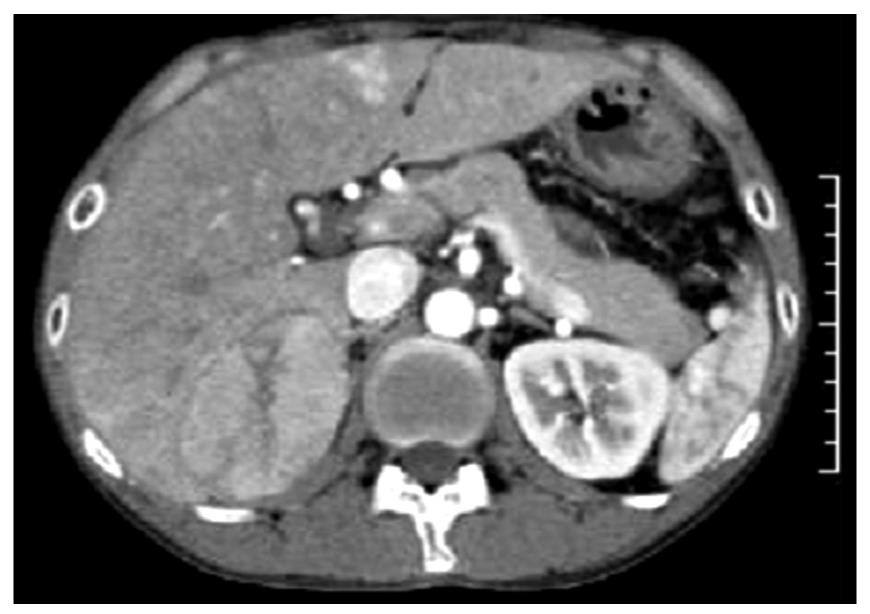
Figure 1 Contrast-enhanced abdominal computed tomography.
A well-circumscribed tumor showing early arterial enhancement is present in S7.
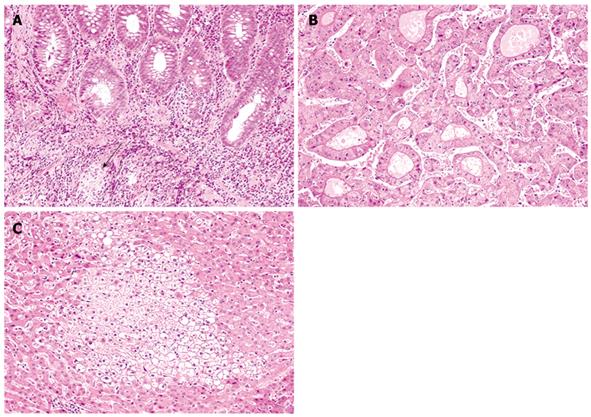
Figure 2 The histopathology of colorectal mucosa (A), the liver tumor (B) and non-neoplastic liver (C) (hematoxylin and eosion stain, × 100).
A: Lymphoplasmacytic infiltrate and a tiny granuloma without association with crypt rupture (arrow) are observed; B: The neoplastic hepatocytes show pseudoglandular growth; C: Focal hepatocyte glycogenosis is observed.
- Citation: Ishida M, Naka S, Shiomi H, Tsujikawa T, Andoh A, Nakahara T, Saito Y, Kurumi Y, Takikita-Suzuki M, Kojima F, Hotta M, Tani T, Fujiyama Y, Okabe H. Hepatocellular carcinoma occurring in a Crohn’s disease patient. World J Gastroenterol 2010; 16(25): 3215-3218
- URL: https://www.wjgnet.com/1007-9327/full/v16/i25/3215.htm
- DOI: https://dx.doi.org/10.3748/wjg.v16.i25.3215


