Copyright
©2010 Baishideng.
World J Gastroenterol. Jul 7, 2010; 16(25): 3206-3210
Published online Jul 7, 2010. doi: 10.3748/wjg.v16.i25.3206
Published online Jul 7, 2010. doi: 10.3748/wjg.v16.i25.3206
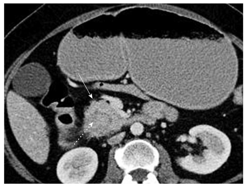
Figure 1 Multislice computed tomography.
A fluid filled stomach and enlargement of the pancreatic head (arrow) are detected, which encircles the second segment of the duodenum (dotted arrow).
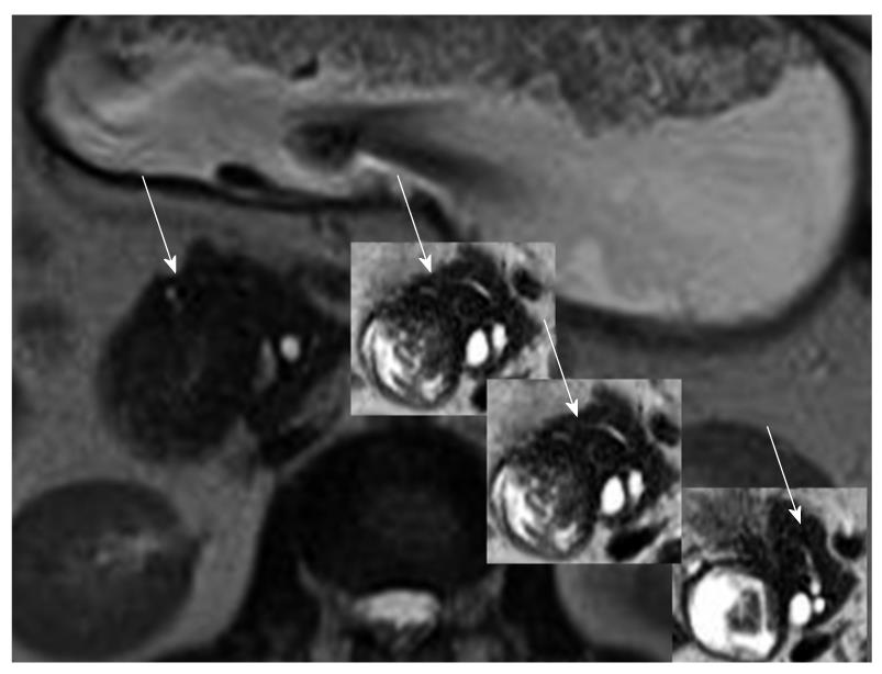
Figure 2 T2 weighted magnetic resonance cholangiopancreatography images depict the aberrant pancreatic duct, which encircles the duodenum and connects with the main pancreatic duct (arrows).
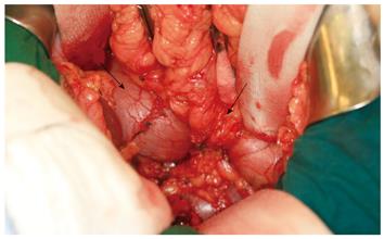
Figure 3 During surgery, a massively distended and elongated first segment (left arrow) and a conic stenosis of the second segment of the duodenum were observed due to the annular pancreas (right arrow).
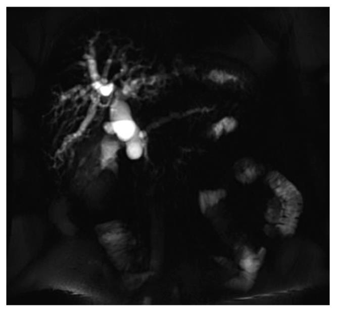
Figure 4 Eight weeks later, T1 w post contrast axial MR shows dilatation of the common bile duct.
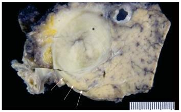
Figure 5 Cross sectional view showing the tumor within the duodenum (asterisk).
The duodenum is surrounded by the incomplete annular pancreas (arrows).
- Citation: Brönnimann E, Potthast S, Vlajnic T, Oertli D, Heizmann O. Annular pancreas associated with duodenal carcinoma. World J Gastroenterol 2010; 16(25): 3206-3210
- URL: https://www.wjgnet.com/1007-9327/full/v16/i25/3206.htm
- DOI: https://dx.doi.org/10.3748/wjg.v16.i25.3206


