Copyright
©2010 Baishideng.
World J Gastroenterol. Jul 7, 2010; 16(25): 3103-3111
Published online Jul 7, 2010. doi: 10.3748/wjg.v16.i25.3103
Published online Jul 7, 2010. doi: 10.3748/wjg.v16.i25.3103
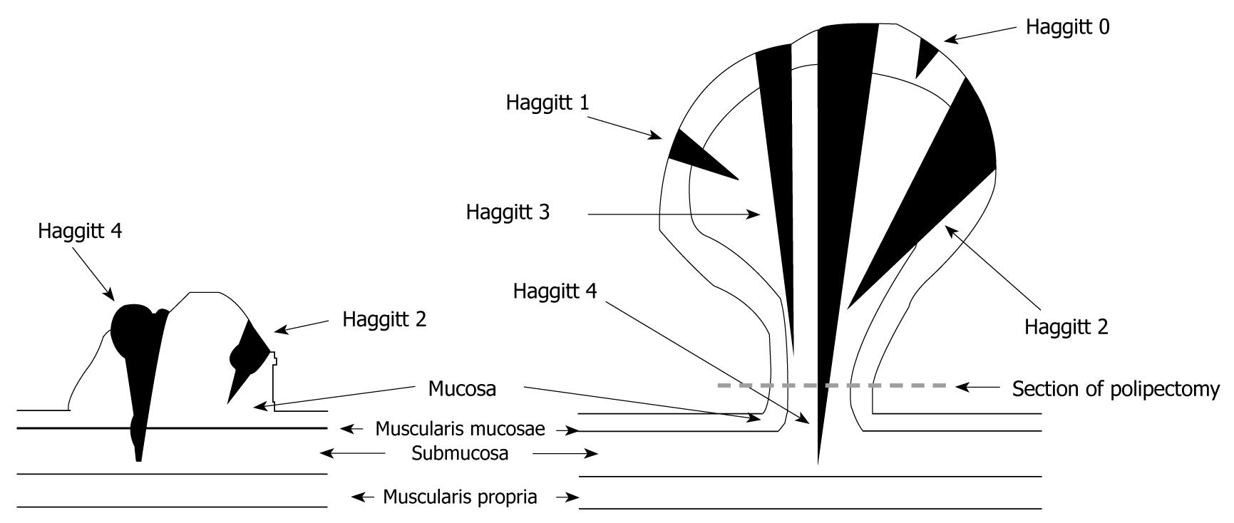
Figure 1 The Haggitt classification.
Haggitt 0: Limited to the mucosa without invasion of submucosa; Haggitt 1: Invasion of submucosa but limited to the head of the polyp; Haggitt 2: Invasion extending into the neck of the polyp; Haggitt 3: Invasion into any part of the stalk; Haggitt 4: Invasion beyond the stalk but above the muscularis propria.
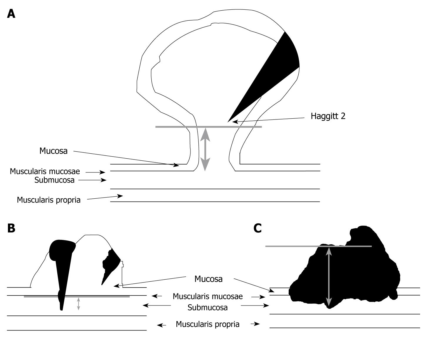
Figure 2 Kitajima’s classification.
The method used for measurement of submucosal invasion (SM) depth. A: For pedunculated submucosal invasive colorectal carcinoma, level 2 according to Haggitt’s classification was used as the baseline (grey line), and SM depth was measured as the vertical distance from this line to the deepest portion of invasion; B: For nonpedunculated, when the muscularis mucosae could be identified in HE stained specimens, the muscularis mucosae was used as baseline (grey line) and the vertical distance from this line to the deepest portion of invasion represented with bidirectional arrow grey; C: For nonpedunculated, when the muscularis mucosae could not be identified due to carcinomatous invasion, the superficial aspect of the invasive carcinoma was used as baseline (grey line), and the vertical distance from this line to the deepest portion of invasion was determined (bidirectional arrow grey).
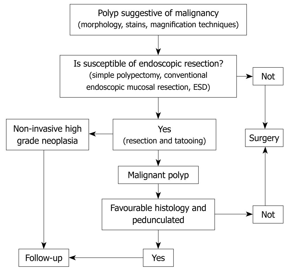
Figure 3 Diagnostic and therapeutic algorithm of malignant polyps.
- Citation: Bujanda L, Cosme A, Gil I, Arenas-Mirave JI. Malignant colorectal polyps. World J Gastroenterol 2010; 16(25): 3103-3111
- URL: https://www.wjgnet.com/1007-9327/full/v16/i25/3103.htm
- DOI: https://dx.doi.org/10.3748/wjg.v16.i25.3103


