Copyright
©2010 Baishideng.
World J Gastroenterol. Jun 28, 2010; 16(24): 3087-3090
Published online Jun 28, 2010. doi: 10.3748/wjg.v16.i24.3087
Published online Jun 28, 2010. doi: 10.3748/wjg.v16.i24.3087
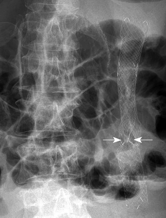
Figure 1 Plain film of the abdomen seven days after deploying the second self-expanding metal stent (SEMS).
The unexpanded part of the second SEMS due to entanglement (arrows).
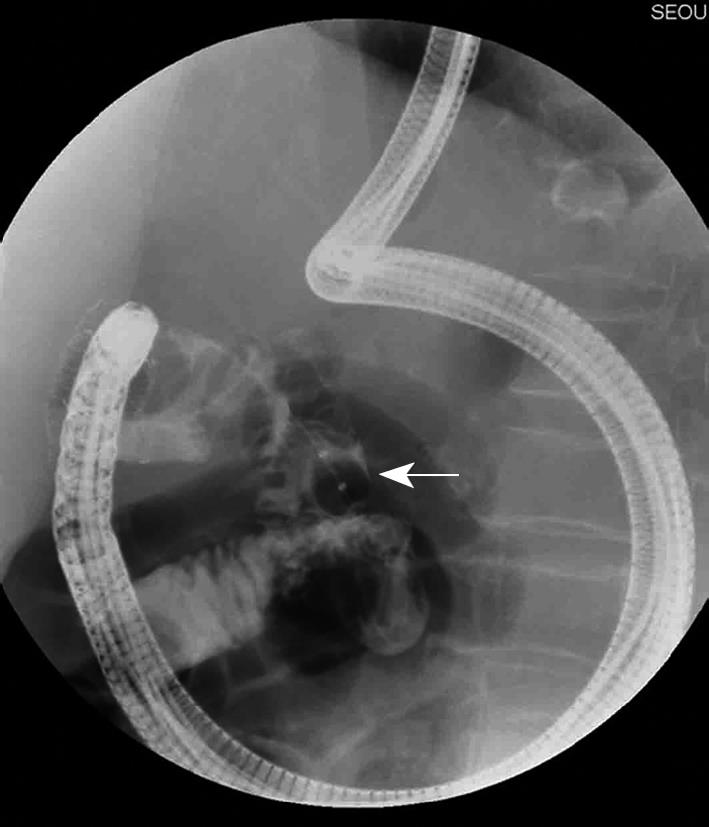
Figure 2 Fluoroscopy.
The stone extraction balloon (arrow) was advanced distal to the existing SEMS.
Figure 3 Duodenoscopy.
A: The stone extraction balloon advancing over the region of tumor ingrowth; B: The guidewire advancing through the stone extraction balloon; C: The expanded covered SEMS.
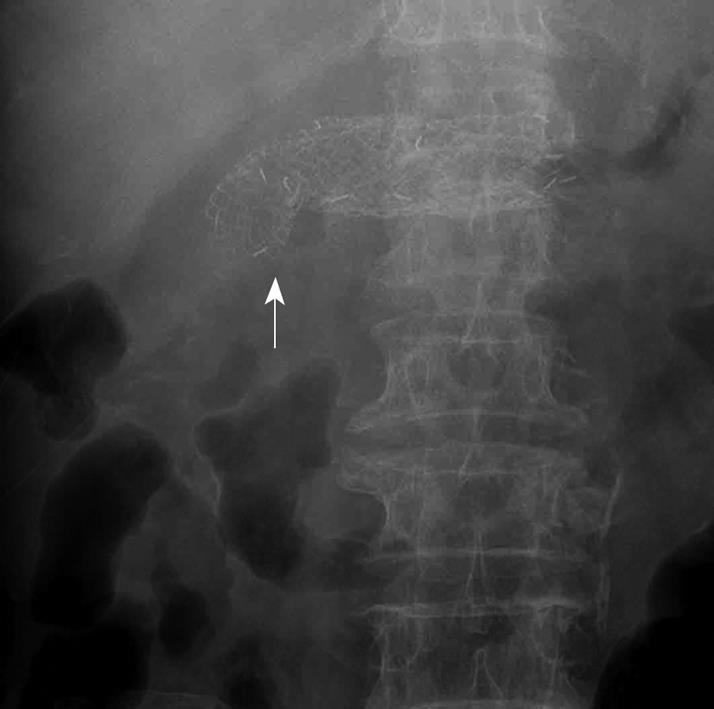
Figure 4 Plain film of the abdomen seven days after replacement of the SEMS.
The fully expanded second SEMS overlapping the first SEMS without entanglement (arrow).
- Citation: Kim HH, Moon JS, Ryu SH, Lee JH, Kim YS. Stone extraction balloon-guided repeat self-expanding metal stent placement. World J Gastroenterol 2010; 16(24): 3087-3090
- URL: https://www.wjgnet.com/1007-9327/full/v16/i24/3087.htm
- DOI: https://dx.doi.org/10.3748/wjg.v16.i24.3087


