Copyright
©2010 Baishideng.
World J Gastroenterol. Jun 14, 2010; 16(22): 2828-2831
Published online Jun 14, 2010. doi: 10.3748/wjg.v16.i22.2828
Published online Jun 14, 2010. doi: 10.3748/wjg.v16.i22.2828
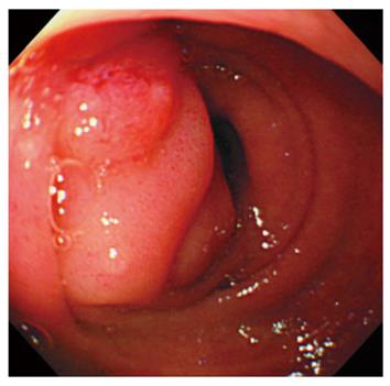
Figure 1 Endoscopic view of the forward-viewing endoscope showing duodenal stricture at the second portion of duodenum.
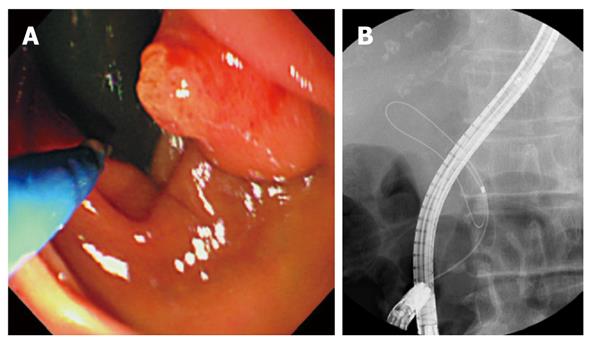
Figure 2 Selective biliary cannulation conducted with a thin forward-viewing endoscope.
A: Endoscopic view showing endoscopic retrograde cholangiopancreatography (ERCP) catheter approaching to the papilla; B: X-ray picture showing common bile duct cannulation conducted with wire-guided technique without contrast injection.
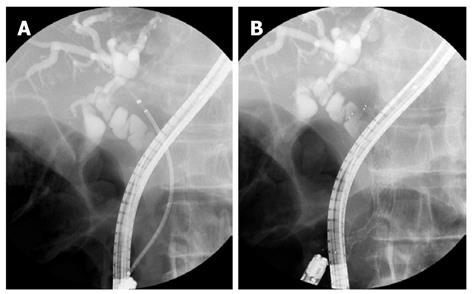
Figure 3 Biliary self-expandable metallic stent (SEMS) placement via a thin forward-viewing endoscope.
A: An extra slim delivery catheter is inserted properly into the bile duct; B: SEMS is successfully placed.
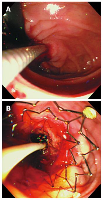
Figure 4 Endoscopic view showing biliary metallic stent placement via a thin forward-viewing endoscope.
A: The delivery catheter is successfully introduced into the bile duct; B: SEMS is successfully placed.
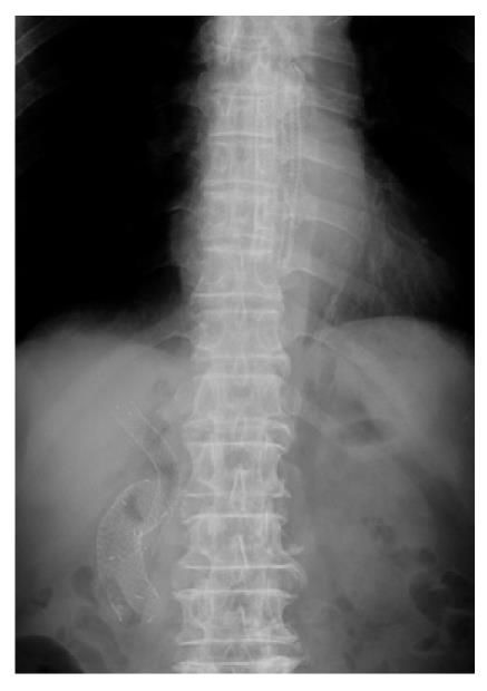
Figure 5 X-ray picture indicating successful placement of biliary, duodenal and esophageal stents.
- Citation: Maetani I, Nambu T, Omuta S, Ukita T, Shigoka H. Treating bilio-duodenal obstruction: Combining new endoscopic technique with 6 Fr stent introducer. World J Gastroenterol 2010; 16(22): 2828-2831
- URL: https://www.wjgnet.com/1007-9327/full/v16/i22/2828.htm
- DOI: https://dx.doi.org/10.3748/wjg.v16.i22.2828


