Copyright
©2010 Baishideng.
World J Gastroenterol. Jun 14, 2010; 16(22): 2735-2742
Published online Jun 14, 2010. doi: 10.3748/wjg.v16.i22.2735
Published online Jun 14, 2010. doi: 10.3748/wjg.v16.i22.2735
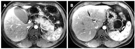
Figure 1 43-year-old woman with acute pancreatitis.
Contrast-enhanced axial T1-weighted images during arterial phase (A) and late venous phase (B, C) show spotted, patchy non-enhanced areas in the pancreatic head and body (arrows).
Figure 2 65-year-old man with acute pancreatitis.
Axial T2-weighted image (A) shows the high-signal intensity in the pancreatic head and body (arrows). The arterial phase MR image (B) shows a large necrotic area (“black pancreas”) in the pancreatic head and body (arrows).
Figure 3 34-year-old man with acute pancreatitis.
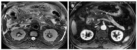
Contrast-enhanced axial T1-weighted images show peripancreatic inflammation extension (arrows) along the mesenteric adipose tissue region (A, B). There is addition irregular thickening and enhancement of the adjacent intestinal wall (arrows, C).
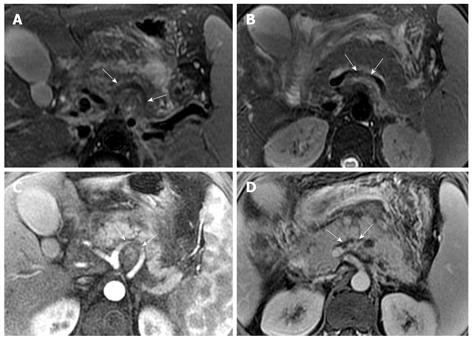
Figure 4 31-year-old man with acute pancreatitis.
Contrast-enhanced axial T1-weighted images with fat suppression reveal thickening and enhancement of dorsal peritoneum (arrow, A). The peripancreatic inflammation extended from the retroperitoneal space to the anterior pararenal space of the left kidney (arrows, B and C).
Figure 5 30-year-old man with acute pancreatitis.
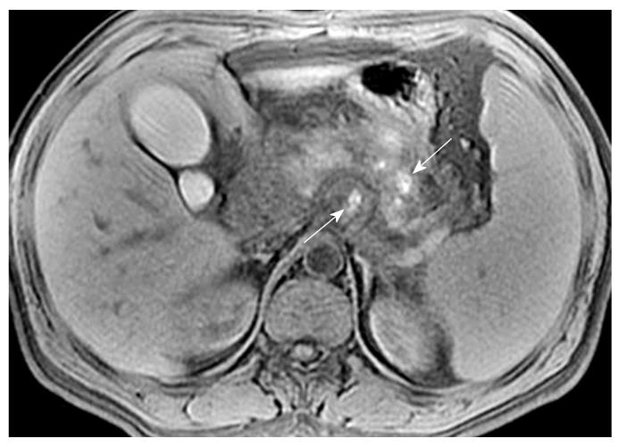
Unenhanced axial T1-weighted image with fat suppression shows patchy hemorrhagic foci (arrows) in the pancreas and in the fatty tissue behind the pancreas.
Figure 6 50-year-old man with acute pancreatitis.
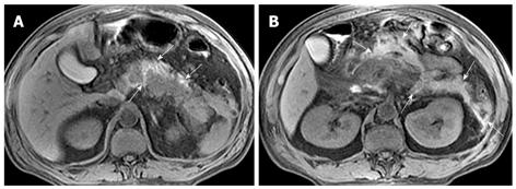
Unenhanced axial T1-weighted images with fat suppression reveal pancreatic and peripancreatic hemorrhage (arrows, A and B); Peripancreatic hemorrhage involvement include the retroperitoneal space and anterior pararenal space of the left kidney (arrows, B).
Figure 7 39-year-old man with acute necrotizing pancreatitis and peripancreatic cellulitis.
Contrast-enhanced T1-weighted images show a ring-like and separated enhancing mass in pancreatic adjacent retroperitoneal space (arrows, A and B).
Figure 8 33-year-old man with acute necrotizing pancreatitis and peripancreatic abscess.
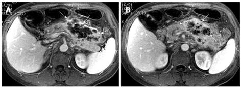
Contrast-enhanced axial T1-weighted images reveal abnormal peripancreatic enhancement (arrows, A) and a liquid-like mass with a well-defined, thickened, and enhancing wall (arrows, B).
Figure 9 41-year-old man with a large peripancreatic pseudocyst who presented as abdominal distension for follow-up after acute pancreatitis.
Unenhanced axial T1-weighted (A) and axial T2-weighted (B) images show a large lesion with typical liquid signal intensity around the pancreas (arrows). MRCP image (C) reveals the pancreas wrapped inside the large pseudocyst, just like the swimming fish (arrow).
Figure 10 35-year-old man with multiple pseudocysts complicated hemorrhage.
Patient had previously suffered from acute pancreatitis. Unenhanced axial T1-weighted images (A and B) with fat suppression and axial T2-weighted image (C) with fat suppression reveal hemorrhagic pseudocysts with a mixed signal intensity dominated by hyperintensity (arrows).
Figure 11 30-year-old man with acute pancreatitis and splenomegaly.
Axial T2-weighted images (A and B) show the involved segments (arrows) of splenic artery (A) and splenic vein (B). Enhanced arterial phase (C) and venous phase (D) images reveal clearly the involved segments (arrows) of the splenic artery (C) and vein (D).
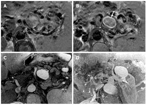
Figure 12 36-year-old man with a history of acute pancreatitis and a splenic artery pseudoaneurysm.
The involved segment of the splenic artery (arrow, A) and an aneurysmal dilatation structure (large arrow, B) are seen on axial T2-weighted images. On enhanced arterial phase image (C), the enhancement of the pseudoaneurysm cavity (white arrow) and the crescent-form filling defect (black arrow) are similar to the Chinese “Yin-Yang” diagram. This pseudoaneurysm is also illustrated on contrast-enhanced MR angiography (arrow, D).
- Citation: Xiao B, Zhang XM, Tang W, Zeng NL, Zhai ZH. Magnetic resonance imaging for local complications of acute pancreatitis: A pictorial review. World J Gastroenterol 2010; 16(22): 2735-2742
- URL: https://www.wjgnet.com/1007-9327/full/v16/i22/2735.htm
- DOI: https://dx.doi.org/10.3748/wjg.v16.i22.2735


