Copyright
©2010 Baishideng.
World J Gastroenterol. Jun 7, 2010; 16(21): 2677-2681
Published online Jun 7, 2010. doi: 10.3748/wjg.v16.i21.2677
Published online Jun 7, 2010. doi: 10.3748/wjg.v16.i21.2677
Figure 1 A lipoma of the ileum in a 56-year-old woman.
A: Intussusception (black arrows) secondary to a lipoma. On transverse enhanced computed tomography (CT) scan, a well-defined hypo-attenuating mass (white arrows) is revealed with no enhancement, but it is difficult to differentiate the tumor from the intraluminal gas (arrowheads); B: The same level image with Figure 1A. It is easy to differentiate the lipoma (black arrows) from the intraluminal gas (white arrows) with regulated image windows; C: Small intestinal two-dimensional (2D) CT enterography shows ileoileac intussusception (white arrows) secondary to a lipoma (black arrows); D: Photomicrograph reveals the lipoma (black arrow) in the submucosal layer, the intestinal normal mucous membrane is also seen (white arrows) (HE, × 40).
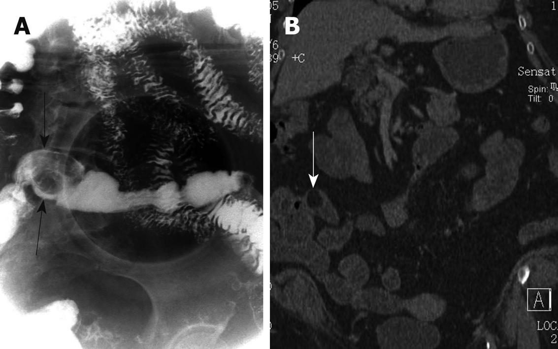
Figure 2 A lipoma of the ileum in a 70-year-old woman.
A: Gastrointestinal barium examination shows an oval well-circumscribed intraluminal mass in the ileum (black arrows); B: Small intestinal 2D CT enterography reveals oval well-circumscribed hypo-intense (-89 HU) intraluminal mass (white arrow) in the ileum.
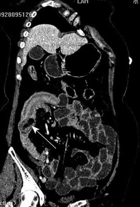
Figure 3 A lipoma of the ileum in a 59-year-old woman.
Small intestinal 2D CT enterography showing intussusception secondary to a lipoma (white arrow).
- Citation: Fang SH, Dong DJ, Chen FH, Jin M, Zhong BS. Small intestinal lipomas: Diagnostic value of multi-slice CT enterography. World J Gastroenterol 2010; 16(21): 2677-2681
- URL: https://www.wjgnet.com/1007-9327/full/v16/i21/2677.htm
- DOI: https://dx.doi.org/10.3748/wjg.v16.i21.2677


