Copyright
©2010 Baishideng.
World J Gastroenterol. Jan 14, 2010; 16(2): 275-278
Published online Jan 14, 2010. doi: 10.3748/wjg.v16.i2.275
Published online Jan 14, 2010. doi: 10.3748/wjg.v16.i2.275
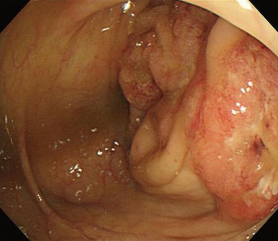
Figure 1 Encircling mass in the ascending colon visualized during colonoscopy.
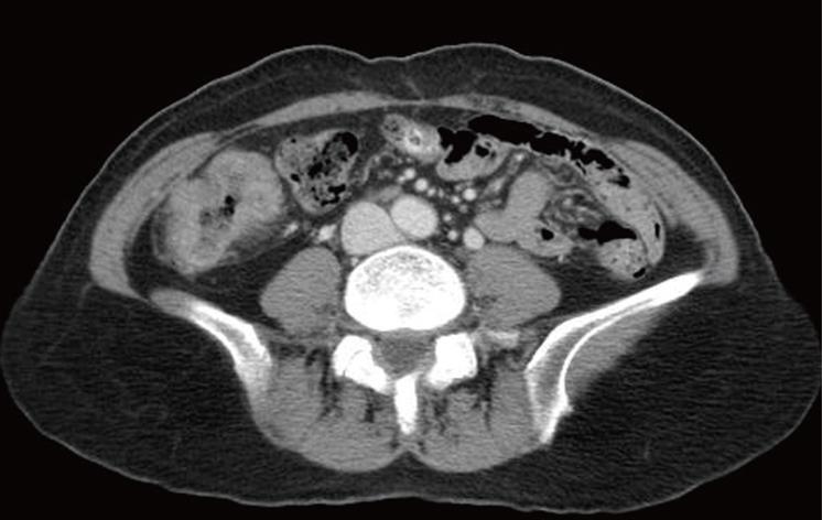
Figure 2 Abdominal CT showed enhanced wall thickening in the ascending colon.
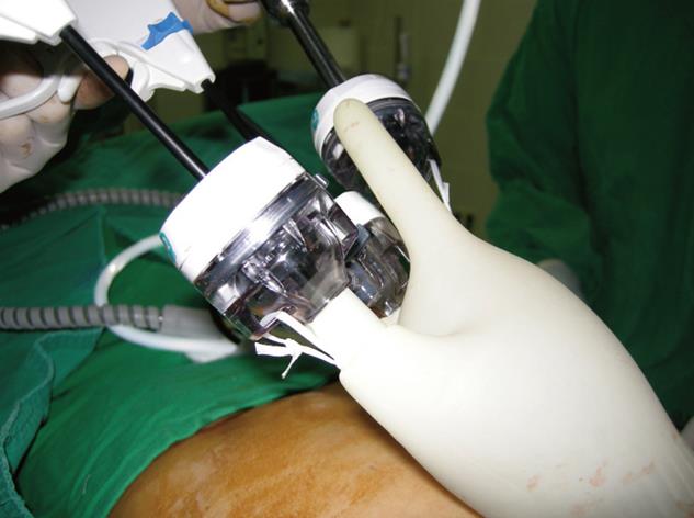
Figure 3 Operative photograph of the single port setting with multiple trocars and instruments.
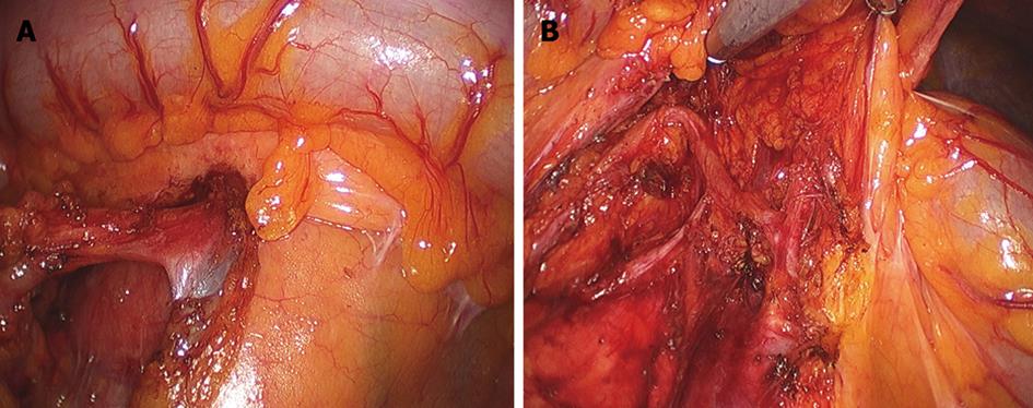
Figure 4 Single port laparoscopic right hemicolectomy with D3 node dissection around the superior mesenteric vein (A) and artery (B).
Figure 5 Surgical wound around the umbilicus (A) and surgical specimen of the resected colon (B and C).
- Citation: Choi SI, Lee KY, Park SJ, Lee SH. Single port laparoscopic right hemicolectomy with D3 dissection for advanced colon cancer. World J Gastroenterol 2010; 16(2): 275-278
- URL: https://www.wjgnet.com/1007-9327/full/v16/i2/275.htm
- DOI: https://dx.doi.org/10.3748/wjg.v16.i2.275


