Copyright
©2010 Baishideng.
World J Gastroenterol. Apr 14, 2010; 16(14): 1808-1810
Published online Apr 14, 2010. doi: 10.3748/wjg.v16.i14.1808
Published online Apr 14, 2010. doi: 10.3748/wjg.v16.i14.1808
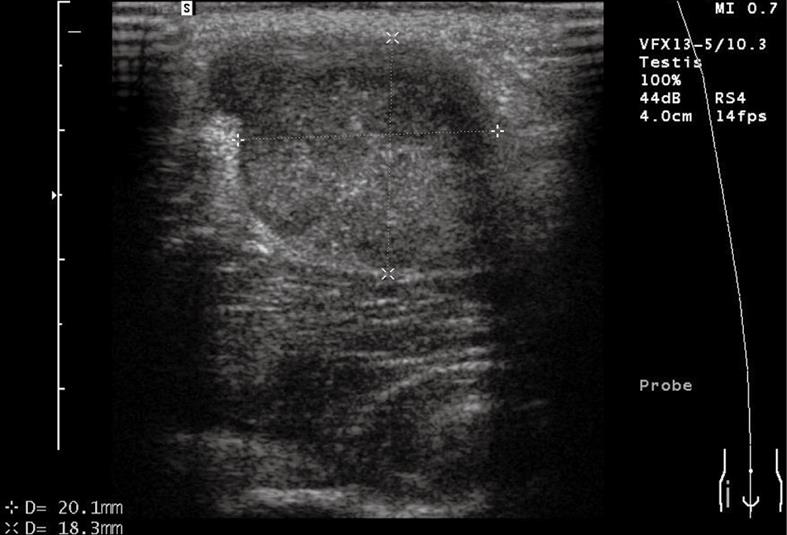
Figure 1 Ultrasonography.
Inguinal ultrasonography shows a low echo-level lesion in the right inguinal region.
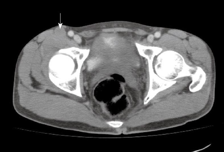
Figure 2 Computed tomography (CT).
A low density lesion with weak, uneven enhancement during contrast-enhanced scan (arrow).
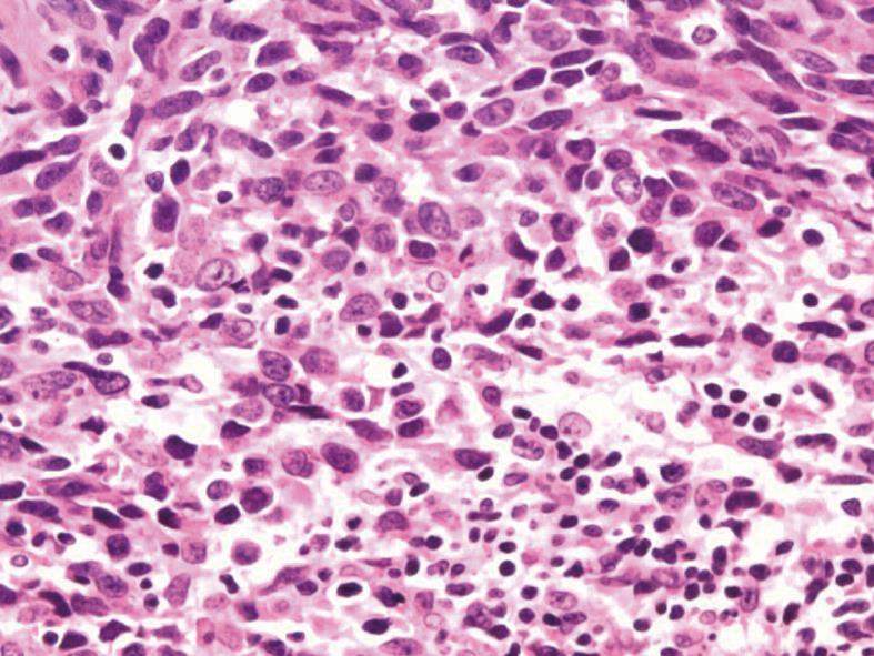
Figure 3 Hematoxylin & eosin (HE) staining.
Spindle cells were predominantly arranged in interweaving fascicles (HE stain, × 100).
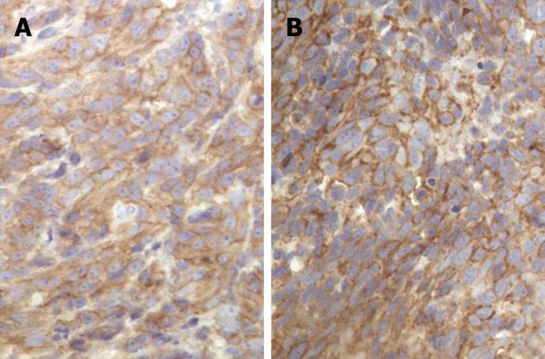
Figure 4 Immunohistochemical staining.
A strong positivity for both CD117 (A) and CD34 (B) (CD117 stain, × 400, CD34 stain, × 400).
- Citation: Zhang Q, Yu JW, Yang WL, Liu XS, Yu JR. Gastrointestinal stromal tumor of stomach with inguinal lymph nodes metastasis: A case report. World J Gastroenterol 2010; 16(14): 1808-1810
- URL: https://www.wjgnet.com/1007-9327/full/v16/i14/1808.htm
- DOI: https://dx.doi.org/10.3748/wjg.v16.i14.1808


