Copyright
©2010 Baishideng.
World J Gastroenterol. Apr 14, 2010; 16(14): 1800-1803
Published online Apr 14, 2010. doi: 10.3748/wjg.v16.i14.1800
Published online Apr 14, 2010. doi: 10.3748/wjg.v16.i14.1800
Figure 1 A huge tumor occupying a wide area of the right lobe.
A: Abdominal computed tomography (CT) showing a pre-treatment dominant lesion located widely in the right lobe (asterisk); B: The tumor (asterisk) has extended contact with the gastrointestinal tract; C: Intraoperatively, Gore-Tex sheets (arrowheads) maintained a space between the tumor (asterisk) and the gastrointestinal tract (under the fingertips); D: Post-operative abdominal CT showing the spacer (arrowheads) around the tumor (asterisk); the spacer maintained a sufficient open space between the tumor and the gastrointestinal tract.
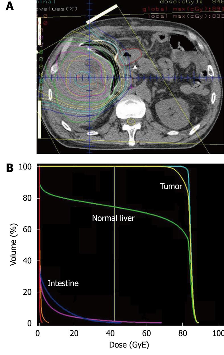
Figure 2 Treatment plan for proton radiotherapy with a total dose of 84 GyE in 20 fractions.
A: Isodose curves demonstrate 100% of the prescribed dose at the center and decreasing by 10% of the dose from the inside out; B: Dose-volume histogram of the target volume and the intestine shows that the tumor is entirely irradiated by almost 100% of the prescribed dose, and the intestine is hardly irradiated.
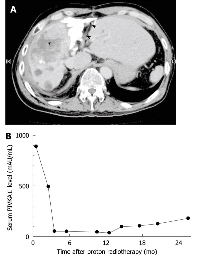
Figure 3 Two years after the proton radiotherapy abdominal CT reveals distinct shrinkage of the tumor (asterisk).
Arrowheads indicate the gastrointestinal tract (A) along with reduced serum levels of the tumor marker, PIVKAII (protein induced by vitamin K absence II) (B).
- Citation: Komatsu S, Hori Y, Fukumoto T, Murakami M, Hishikawa Y, Ku Y. Surgical spacer placement and proton radiotherapy for unresectable hepatocellular carcinoma. World J Gastroenterol 2010; 16(14): 1800-1803
- URL: https://www.wjgnet.com/1007-9327/full/v16/i14/1800.htm
- DOI: https://dx.doi.org/10.3748/wjg.v16.i14.1800


