Copyright
©2010 Baishideng.
World J Gastroenterol. Apr 14, 2010; 16(14): 1735-1741
Published online Apr 14, 2010. doi: 10.3748/wjg.v16.i14.1735
Published online Apr 14, 2010. doi: 10.3748/wjg.v16.i14.1735
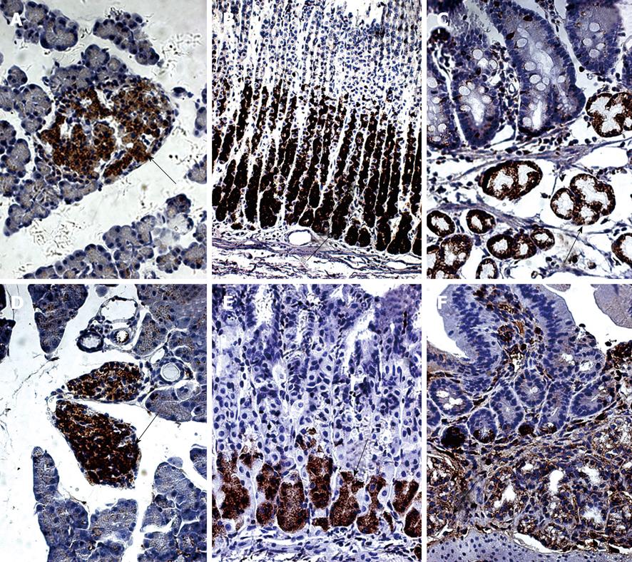
Figure 1 Immunolocalization of nesfatin-1/nucleobindin-2 (NUCB2) in the pancreas, stomach and duodenum in sprague-dawley (SD) rats (A-C) and institute of Cancer Research (ICR) mice (D-F).
A and D: Pancreas; B and E: Stomach; C and F: Duodenum. Tissues were subjected to immunohistochemistry using antibodies to nesfatin-1. Arrows denote areas of positivity (A, C-F: × 40; B: × 20).
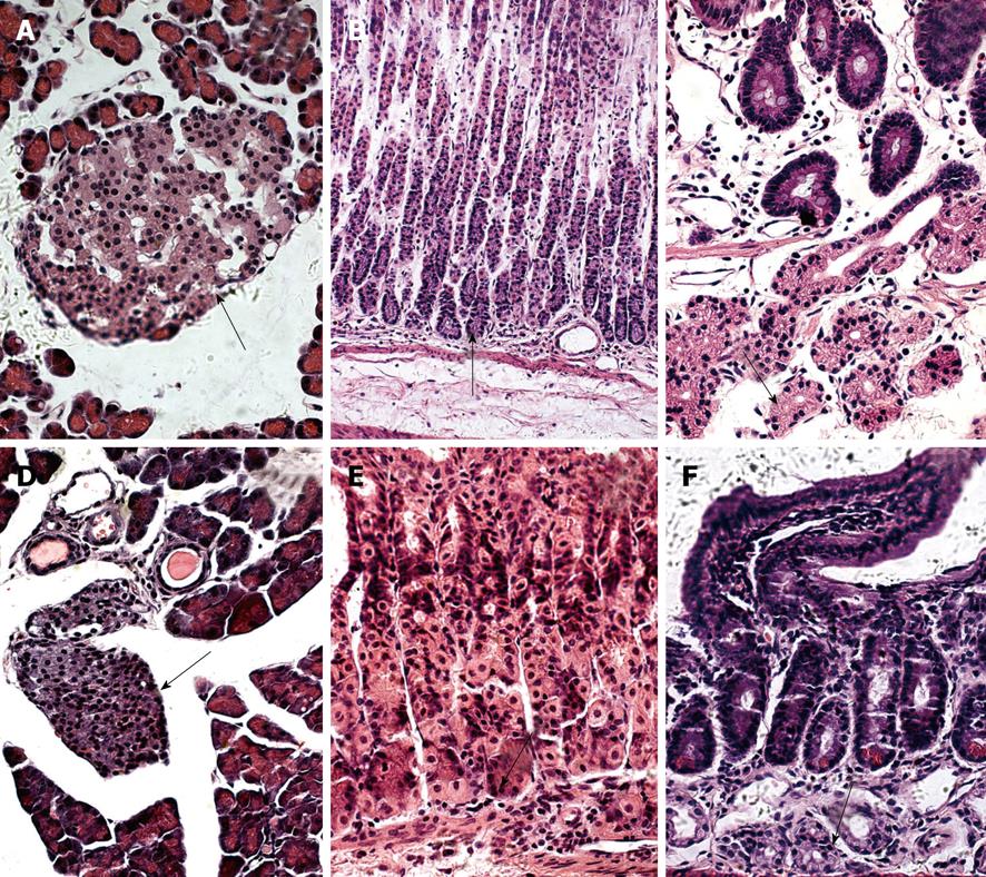
Figure 2 Representative microphotographs of nesfatin-1/NUCB2 in the pancreas, stomach and duodenum in SD rats (A-C) and ICR mice (D-F).
A and D: Pancreas; B and E: Stomach; C and F: Duodenum. Tissues were subjected to hematoxylin and eosin (HE) staining. Arrows denote areas of the same field of view as the immunohistochemistry positivity (A, C-F: × 40; B: × 20).
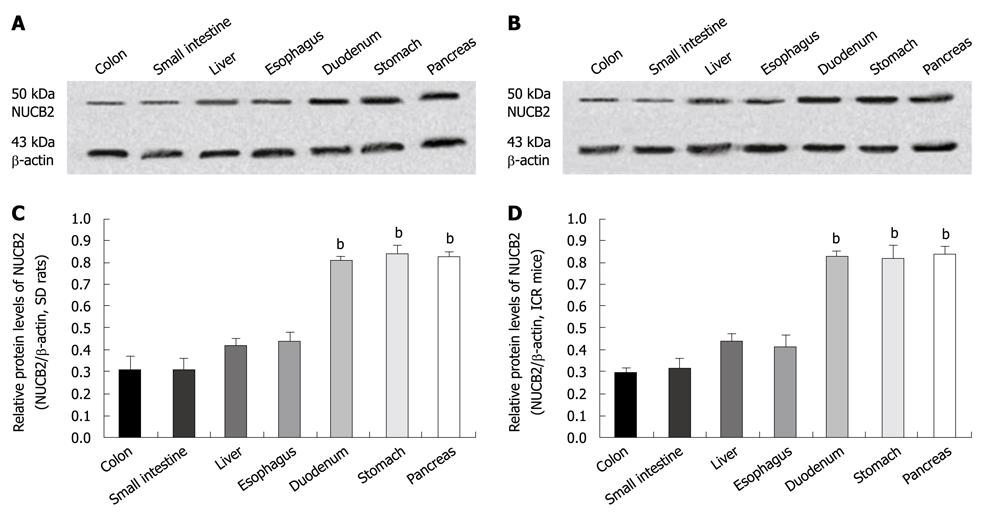
Figure 3 Western blotting for NUCB2 protein in the digestive system of the SD rats (A, C) and ICR mice (B, D).
A, B: The predicted band for full-length NUCB2 protein
- Citation: Zhang AQ, Li XL, Jiang CY, Lin L, Shi RH, Chen JD, Oomura Y. Expression of nesfatin-1/NUCB2 in rodent digestive system. World J Gastroenterol 2010; 16(14): 1735-1741
- URL: https://www.wjgnet.com/1007-9327/full/v16/i14/1735.htm
- DOI: https://dx.doi.org/10.3748/wjg.v16.i14.1735


