Copyright
©2010 Baishideng.
World J Gastroenterol. Apr 7, 2010; 16(13): 1676-1679
Published online Apr 7, 2010. doi: 10.3748/wjg.v16.i13.1676
Published online Apr 7, 2010. doi: 10.3748/wjg.v16.i13.1676
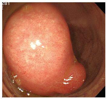
Figure 1 Colonoscopy showing a large pedunculated tumor, 50 mm in size, originating from the ileum end.
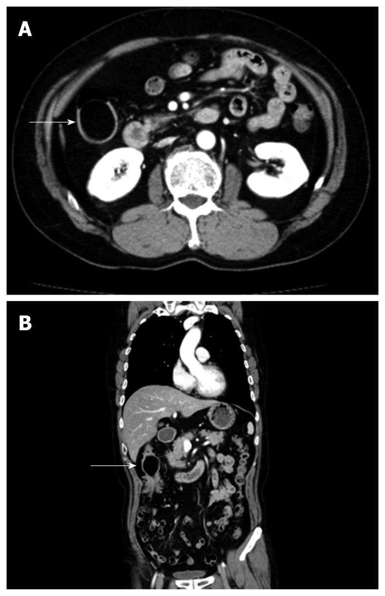
Figure 2 Abdominal CT showing a round, smooth and well-demarcated tumor at the end of the ileum (A) with a fat attenuation coefficient of -116 Hounsfield units (arrows) (B).
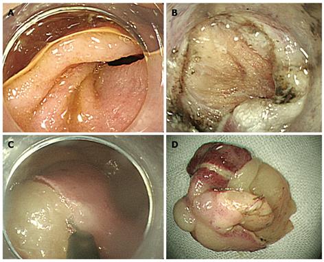
Figure 3 Whole captured lesion after injection of glycerol at the base of the lesion.
The base of the lipoma (A), lacerated muscle layer (B), dissected overlying mucosa and capsule (C), and completely removed lipoma (D).
- Citation: Morimoto T, Fu KI, Konuma H, Izumi Y, Matsuyama S, Ogura K, Miyazaki A, Watanabe S. Peeling a giant ileal lipoma with endoscopic unroofing and submucosal dissection. World J Gastroenterol 2010; 16(13): 1676-1679
- URL: https://www.wjgnet.com/1007-9327/full/v16/i13/1676.htm
- DOI: https://dx.doi.org/10.3748/wjg.v16.i13.1676


