Copyright
©2010 Baishideng.
World J Gastroenterol. Apr 7, 2010; 16(13): 1660-1664
Published online Apr 7, 2010. doi: 10.3748/wjg.v16.i13.1660
Published online Apr 7, 2010. doi: 10.3748/wjg.v16.i13.1660
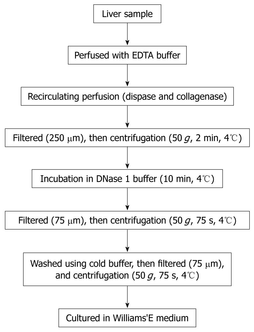
Figure 1 Flow diagram of the preparation of isolated hepatocytes with a modified four-step retrograde perfusion technique.
Figure 2 Isolation technique and microscopic appearance of primary and immortalized porcine hepatocytes.
A: Perfusion was conducted by inserting a suitable pipette into vessels exposed on a cut surface of the sample; B: Non-immortalized primary porcine hepatocytes grow slowly with low plating efficiency and a short life span; C: Immortalized porcine hepatocytes grow rapidly as islands and had an extended life span. Magnifications: B and C 100 ×.
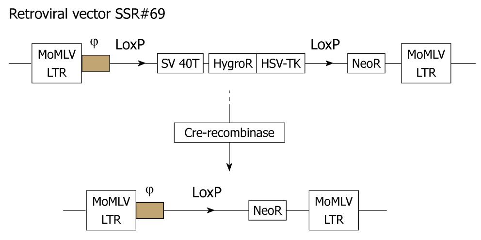
Figure 3 Schematic drawings of the integrating component of retroviral vector SSR#69 before and after Cre-recombination.
SSR#69 contains the hygromycin B resistance gene (Hyg R) as a positive selectable marker and the herpes simplex virus thymidine kinase gene (HSV-TK) as a negative selectable marker. The SV40T, Hyg R and HSV-TK genes are flanked by loxP sites.
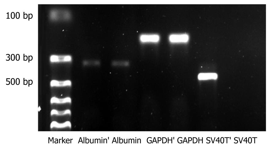
Figure 4 Gene expression of liver-specific functions in immortalized and reverted cells.
Lines 1 to 7, from left to right, Marker, Albumin’ (immortalized hepatocytes), Albumin (reverted hepatocytes), GAPDH’ (immortalized hepatocytes), GAPDH (reverted hepatocytes), SV40T’ (immortalized hepatocytes), SV40T (reverted hepatocytes), respectively.
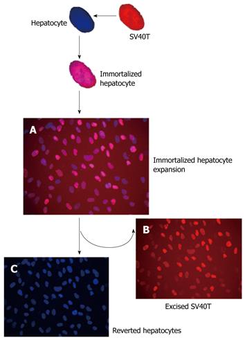
Figure 5 Scheme of reversible immortalization.
The primary porcine hepatocytes were immortalized by transfer of an oncogene (SV40T). After expansion of the immortalized cells, Cre/loxP recombination was performed to remove the oncogene (SV40T) and the cells reverted to their pre-immortalized state. (A-C) Double immunofluorescence of immortalized hepatocytes stained with DAPI (blue), which binds, together with monoclonal antibody anti-SV40T revealed with texas red-antibody conjugate (red). Blue and red fluorescence merged as purple. SV40T expression is revealed by intense staining of the cell nucleus.
- Citation: Meng FY, Chen ZS, Han M, Hu XP, He XX, Liu Y, He WT, Huang W, Guo H, Zhou P. Porcine hepatocyte isolation and reversible immortalization mediated by retroviral transfer and site-specific recombination. World J Gastroenterol 2010; 16(13): 1660-1664
- URL: https://www.wjgnet.com/1007-9327/full/v16/i13/1660.htm
- DOI: https://dx.doi.org/10.3748/wjg.v16.i13.1660


