Copyright
©2010 Baishideng.
World J Gastroenterol. Jan 7, 2010; 16(1): 42-47
Published online Jan 7, 2010. doi: 10.3748/wjg.v16.i1.42
Published online Jan 7, 2010. doi: 10.3748/wjg.v16.i1.42
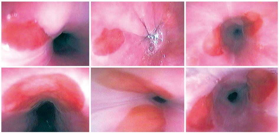
Figure 1 Various endoscopic images of inlet patches (single or double).
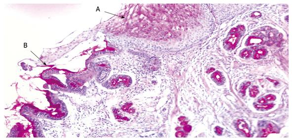
Figure 2 Biopsies.
A shows heterotopic gastric mucosa, B shows squamous epithelium.
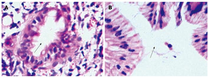
Figure 3 The presence of Hp bacilli in heterotopic gastric mucosa with HE stain (× 1000).
A: Few Hp bacilli (arrow); B: Hp colonization (arrow).
-
Citation: Alagozlu H, Simsek Z, Unal S, Cindoruk M, Dumlu S, Dursun A. Is there an association between
Helicobacter pylori in the inlet patch and globus sensation? World J Gastroenterol 2010; 16(1): 42-47 - URL: https://www.wjgnet.com/1007-9327/full/v16/i1/42.htm
- DOI: https://dx.doi.org/10.3748/wjg.v16.i1.42


