Copyright
©2010 Baishideng.
World J Gastroenterol. Jan 7, 2010; 16(1): 123-125
Published online Jan 7, 2010. doi: 10.3748/wjg.v16.i1.123
Published online Jan 7, 2010. doi: 10.3748/wjg.v16.i1.123
Figure 1 Endoscopic view.
A: An anastomotic ulcer was observed in the cholecystojejunostomy; B: Anastomotic ulcer after argon plasma coagulation therapy; C: n-butyl-2-cyanoacrylate injection into the cholecystojejunostomy varices.
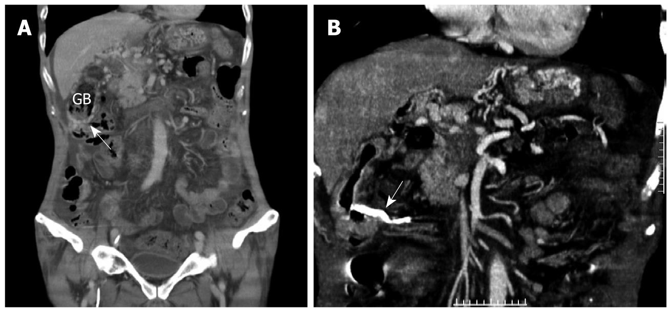
Figure 2 Abdominal computed tomography.
A:Prominent vessels (arrow) formed around the gall bladder; B: Obliterated varicose vein (arrow).
- Citation: Hsu YC, Yen HH, Chen YY, Soon MS. Successful endoscopic sclerotherapy for cholecystojejunostomy variceal bleeding in a patient with pancreatic head cancer. World J Gastroenterol 2010; 16(1): 123-125
- URL: https://www.wjgnet.com/1007-9327/full/v16/i1/123.htm
- DOI: https://dx.doi.org/10.3748/wjg.v16.i1.123


