Copyright
©2009 The WJG Press and Baishideng.
World J Gastroenterol. Feb 21, 2009; 15(7): 888-891
Published online Feb 21, 2009. doi: 10.3748/wjg.15.888
Published online Feb 21, 2009. doi: 10.3748/wjg.15.888
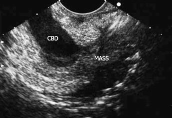
Figure 1 A heterogenic and hypoechoic mass (MASS) with ill-defined margin about 2.
7 cm × 1.8 cm at the junction of the CBD and MPD.
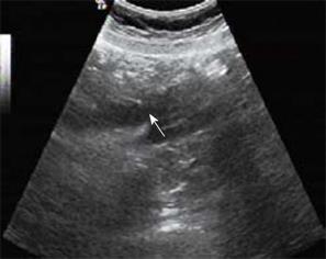
Figure 2 The end part of the CBD (arrow), which suddenly became narrow with a diameter of 0.
7 cm. There was no exact mass detected.
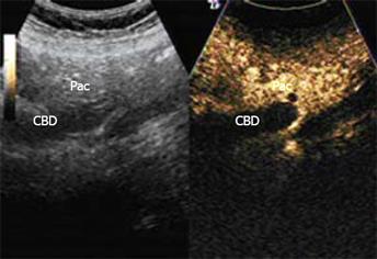
Figure 3 The wall of the CBD enhanced at 12 s after contrast agent was administered, while there was no obvious hyper-enhanced or hypo-enhanced lesion in the ampulla of Vater.
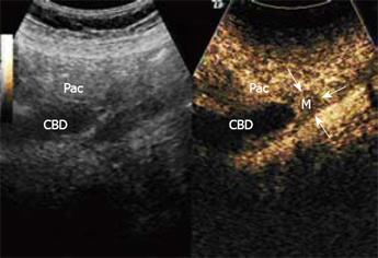
Figure 4 A hypo-enhanced lesion about 1.
7 cm × 1.6 cm with blurred borders in the ampulla of Vater, from 20 s to 180 s, in comparison with the adjacent pancreas Pac.
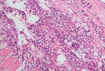
Figure 5 Micrograph shows SRCC of ampulla of Vater (HE × 100).
- Citation: Gao JM, Tang SS, Fu W, Fan R. Signet-ring cell carcinoma of ampulla of Vater: Contrast-enhanced ultrasound findings. World J Gastroenterol 2009; 15(7): 888-891
- URL: https://www.wjgnet.com/1007-9327/full/v15/i7/888.htm
- DOI: https://dx.doi.org/10.3748/wjg.15.888


