Copyright
©2009 The WJG Press and Baishideng.
World J Gastroenterol. Feb 14, 2009; 15(6): 748-752
Published online Feb 14, 2009. doi: 10.3748/wjg.15.748
Published online Feb 14, 2009. doi: 10.3748/wjg.15.748
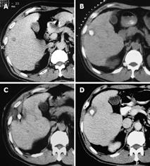
Figure 1 Pathological examinations of biopsied specimens and enhanced CT after PMCT showed complete tumor necrosis in 34 foci, together with complete ring-shaped necrosis of the surrounding non-cancerous hepatic parenchymal tissue, measuring 3-5 mm in width.
A: CT scan of a primary HCC in the right anterior lobe of the liver, with a diameter of 1.6 cm × 1.4 cm; B: After TACE, the accumulation of iodized oil in the tumor area was satisfactory; C: PMCT was initiated 17 d later; D: One month after PMCT, an enhanced CT scan showed a complete non-enhanced area of the tumor, and a non-cancerous ring-shaped area surrounding the tumor (measuring 4-5 mm), indicating complete necrosis of the tumor lesion.
- Citation: Yang WZ, Jiang N, Huang N, Huang JY, Zheng QB, Shen Q. Combined therapy with transcatheter arterial chemoembolization and percutaneous microwave coagulation for small hepatocellular carcinoma. World J Gastroenterol 2009; 15(6): 748-752
- URL: https://www.wjgnet.com/1007-9327/full/v15/i6/748.htm
- DOI: https://dx.doi.org/10.3748/wjg.15.748


