Copyright
©2009 The WJG Press and Baishideng.
World J Gastroenterol. Feb 7, 2009; 15(5): 622-627
Published online Feb 7, 2009. doi: 10.3748/wjg.15.622
Published online Feb 7, 2009. doi: 10.3748/wjg.15.622
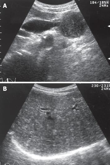
Figure 1 Abdominal US.
A: Pancreatic head mass with hypo-echogenicity, 43 mm in diameter, which compressed the lower portion of the bile duct, and resulted in upstream bile tract dilatation; B: Many tiny, diffuse hyperechoic patches or comet-like tails that suggested the presence of certain intrahepatic abnormalities, but the reason could not be elucidated.
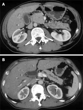
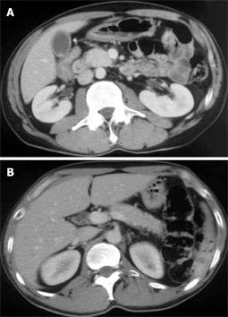
Figure 2 Abdominal enhanced CT imaging with contrast medium in May 2007.
A: Swelling of the pancreatic head like a solid tumor was shown. B: The distal portion of the main pancreatic duct was found to be irregularly dilated; however, the pancreatic body and tail were not atrophied as seen in pancreatic cancer.
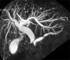
Figure 3 MRCP showed proximal stenosis of the bile duct as well as the main pancreatic duct; however, narrowing of the pancreatic duct, as seen in AIP, was not demonstrated at all.
A simultaneous finding was that there were many round high-intensity nodules scattered throughout the liver.
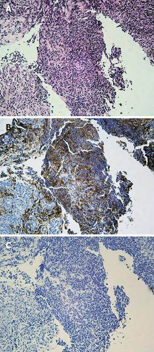
Figure 4 Histopathology of a specimen taken from the pancreatic head mass showed no malignant cells that indicated cancer.
A: Marked infiltration of pancreatic parenchyma by lymphocytic mononuclear cells, as well as interstitial fibrosis suggested a diagnosis of AIP (HE, × 200). B: These infiltrated cells were revealed to be mainly T lymphocytes (T-cell staining, × 200). However, the histopathology was not diagnostic without so-called LPSP with scarce plasma cell infiltration. C: Immunohistochmistry using antibody against IgG4 resulted in a negative study (IgG4 staining, × 200).
Figure 5 Abdominal enhanced CT scan with contrast medium in July 2007.
As compared with CT imaging taken in May 2007, swelling of the pancreatic head had significantly regressed (A). Irregular dilatation of the main pancreatic duct had improved with the whole body bulk being reduced (B).
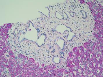
Figure 6 Histopathology of specimens taken from the hepatic parenchyma (PAS, × 200).
Distribution of many irregular, angulated duct structures was observed. Ductal epithelium was comprised of monolayered columnar epithelium, morphologically identical to biliary epithelium, surrounded by fibrous connective tissues with minimal inflammatory change. The sequential histopathological features were considered to be bile duct hamartoma (VMC). Subsequent immunohistochemistry of the region by anti-IgG4 antibody was negative.
- Citation: Miura H, Kitamura S, Yamada H. A variant form of autoimmune pancreatitis successfully treated by steroid therapy, accompanied by von Meyenburg complex. World J Gastroenterol 2009; 15(5): 622-627
- URL: https://www.wjgnet.com/1007-9327/full/v15/i5/622.htm
- DOI: https://dx.doi.org/10.3748/wjg.15.622


