Copyright
©2009 The WJG Press and Baishideng.
World J Gastroenterol. Dec 21, 2009; 15(47): 6007-6009
Published online Dec 21, 2009. doi: 10.3748/wjg.15.6007
Published online Dec 21, 2009. doi: 10.3748/wjg.15.6007
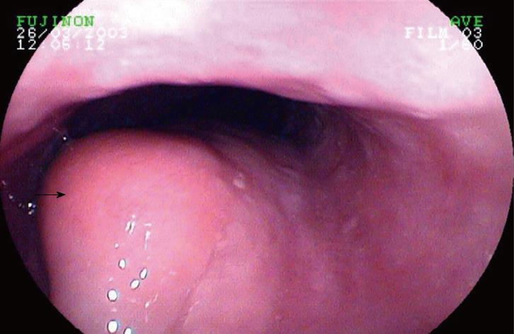
Figure 1 Gastroscopy showing a 4 cm fusiform polyp (black arrow) in mid-esophagus.
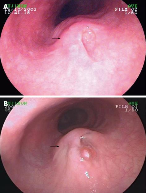
Figure 2 Gastroscopy showing an orifice-like lesion on the top surface of the polyp (black arrows).
A: 1st; B: 2nd.
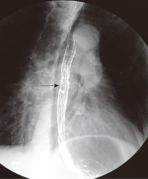
Figure 3 Barium radiography reported a 2 cm filling defect (black arrow) in the mid-esophagus.
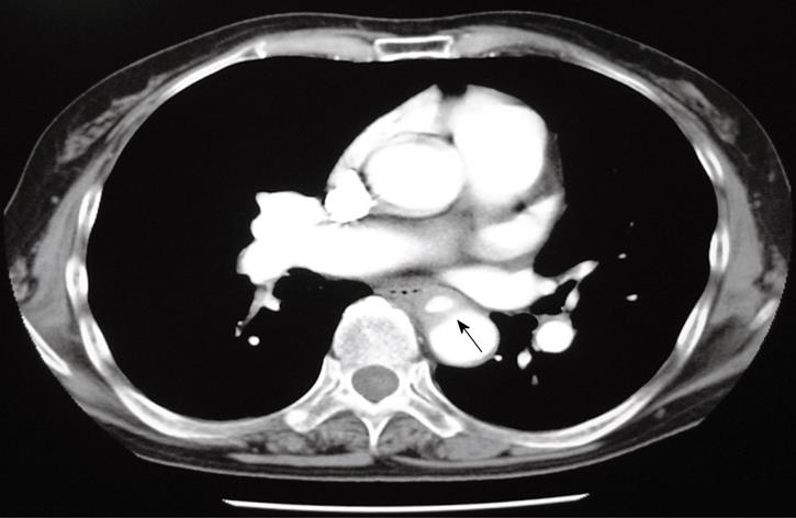
Figure 4 A subsequent enhanced computed tomography (CT) showing a descending aortic pseudoaneurysm (black arrow).
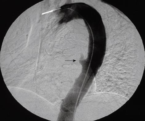
Figure 5 Aortography.
The contrast extravasated from the descending aorta rupture with a diameter of about 0.5 cm (black arrow).
- Citation: Jiao Y, Zong Y, Yu ZL, Yu YZ, Zhang ST. Aortoesophageal fistula: A case misdiagnosed as esophageal polyp. World J Gastroenterol 2009; 15(47): 6007-6009
- URL: https://www.wjgnet.com/1007-9327/full/v15/i47/6007.htm
- DOI: https://dx.doi.org/10.3748/wjg.15.6007


