Copyright
©2009 The WJG Press and Baishideng.
World J Gastroenterol. Dec 21, 2009; 15(47): 6004-6006
Published online Dec 21, 2009. doi: 10.3748/wjg.15.6004
Published online Dec 21, 2009. doi: 10.3748/wjg.15.6004
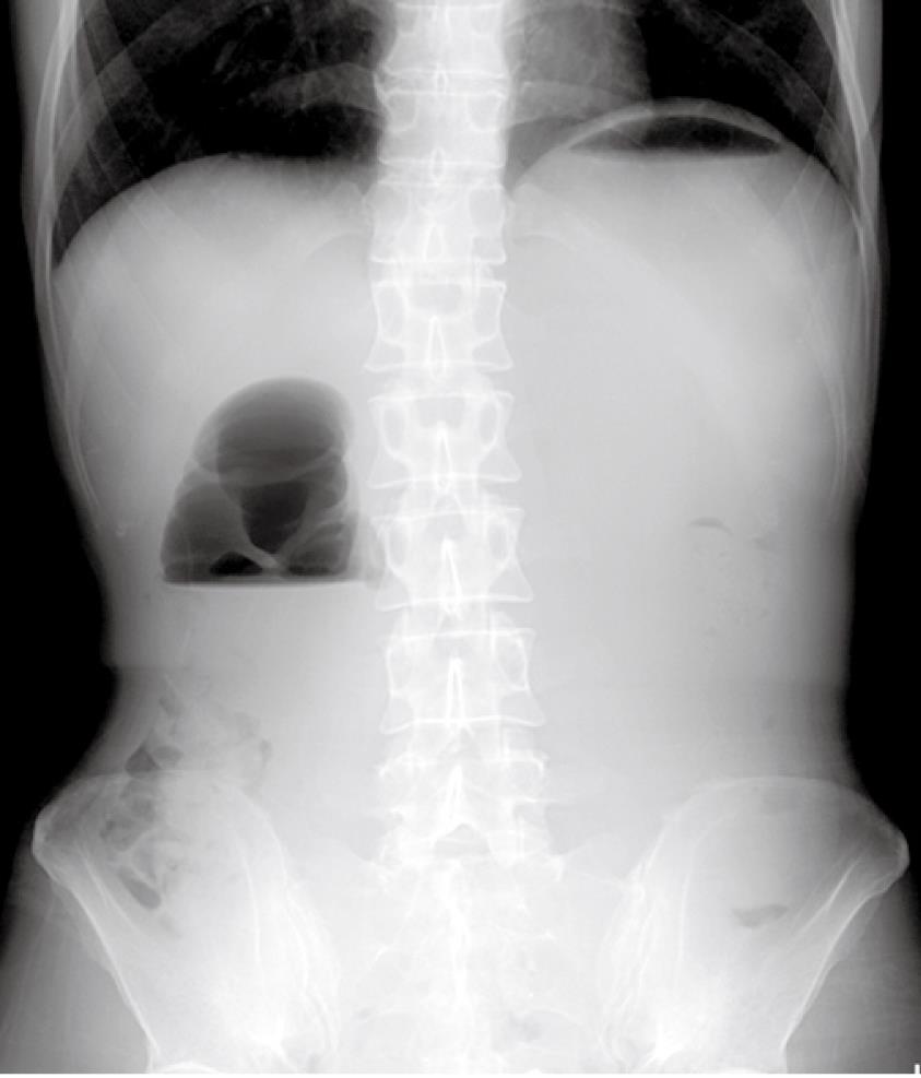
Figure 1 Abdominal X-ray showing a distended stomach with air fluid level in the stomach and duodenal bulb.
The “Double bubble sign” was consistent with high small bowel obstruction.
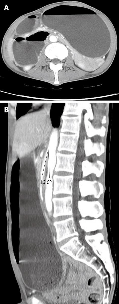
Figure 2 Computed tomography (CT) scan showing distended stomach and 2nd portion of duodenum (A); The angle between aorta and superior mesenteric artery (SMA) was 16.
6° (B).
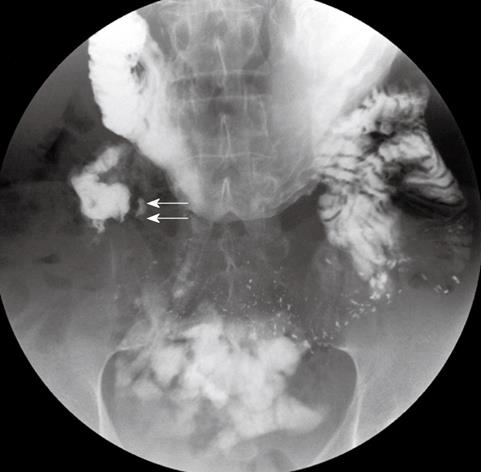
Figure 3 Upper gastrointestinal series showing an abrupt cut-off (short arrows) at the third portion of the duodenum.
- Citation: Wu MC, Wu IC, Wu JY, Wu DC, Wang WM. Superior mesenteric artery syndrome in a diabetic patient with acute weight loss. World J Gastroenterol 2009; 15(47): 6004-6006
- URL: https://www.wjgnet.com/1007-9327/full/v15/i47/6004.htm
- DOI: https://dx.doi.org/10.3748/wjg.15.6004


