Copyright
©2009 The WJG Press and Baishideng.
World J Gastroenterol. Dec 7, 2009; 15(45): 5662-5668
Published online Dec 7, 2009. doi: 10.3748/wjg.15.5662
Published online Dec 7, 2009. doi: 10.3748/wjg.15.5662
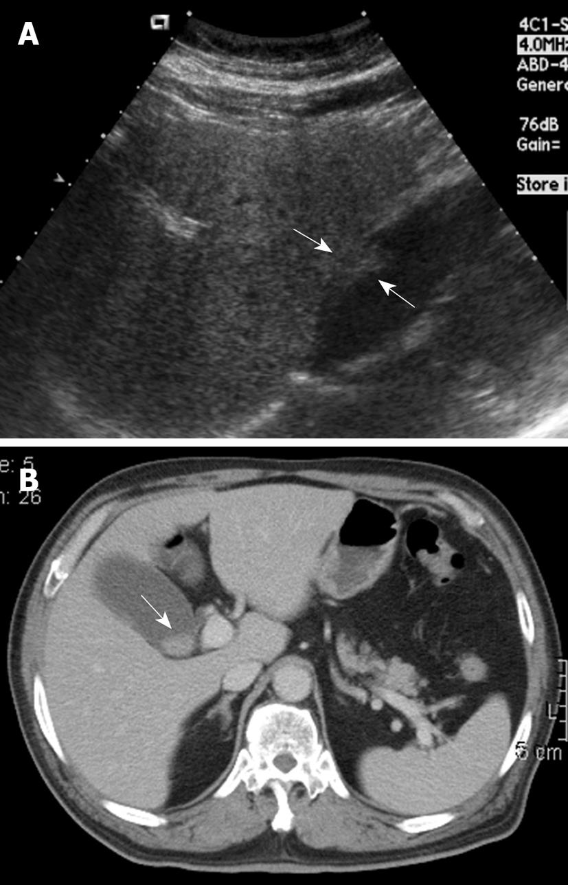
Figure 1 A 68-year-old woman with intermittent dull right upper quadrant pain for 6 mo.
A: Ultrasound (US) showed a sessile polypoid lesion (arrows) with a mildly uneven surface; B: Contrast-enhanced computed tomography (CT) showed an intraluminal polypoid lesion (arrow) with no apparent enlarged regional lymph node.
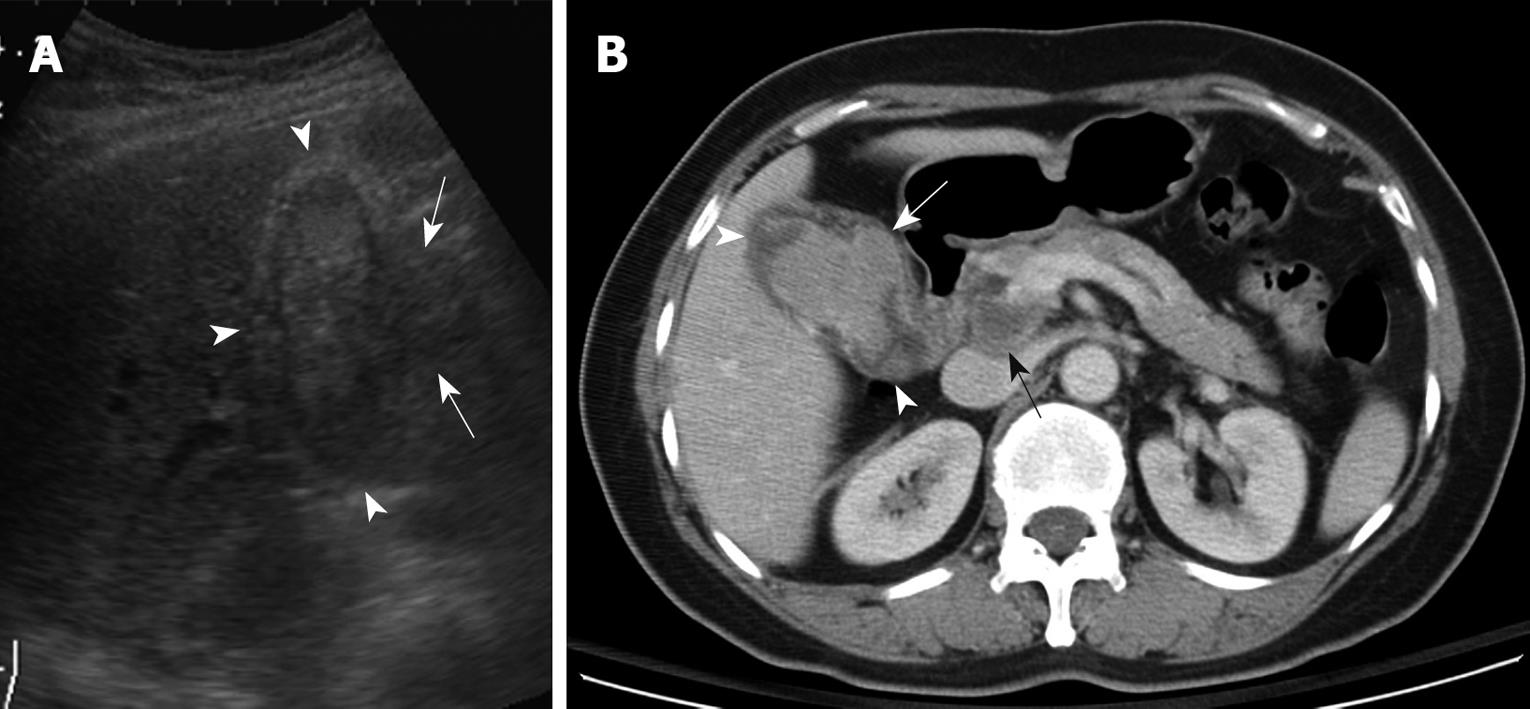
Figure 2 A 78-year-old woman with right upper abdominal pain and progressive jaundice for 2 mo.
A: US showed an intraluminal heteroechoic mass that occupied nearly the whole gallbladder (arrowheads), with focal extraluminal invasion (arrows); B: Contrast-enhanced CT showed a large lobulated mass within the gallbladder (arrowheads), with extracholecystic invasion (white arrow) and hepatoduodenal ligament lymph node metastasis (black arrow).
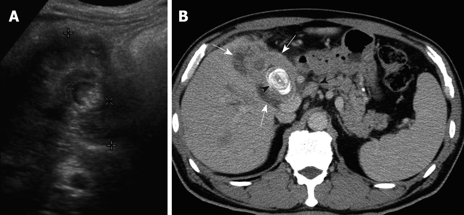
Figure 3 A 77-year-old woman with right upper abdominal pain and jaundice for 1 mo.
A: US showed a large gallstone (× cursors) encased by the diffusedly thickened gallbladder wall, with a heteroechoic appearance (+ cursors); B: Contrast-enhanced CT showed diffuse, uneven wall thickening of the gallbladder (white arrows), with a large laminated gallstone (arrowhead) and hepatoduodenal ligament lymph node metastasis (black arrow).
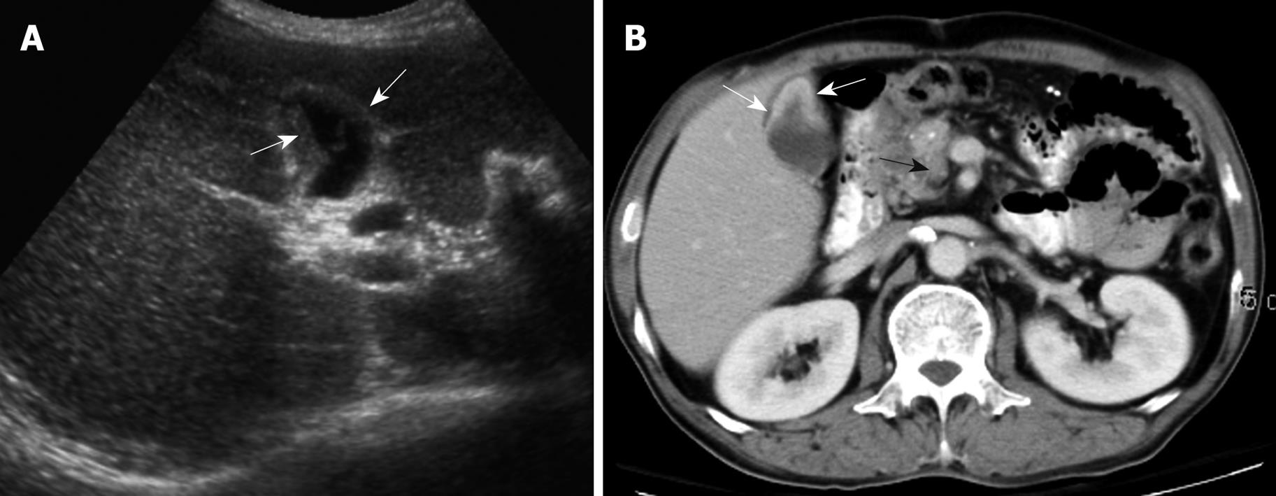
Figure 4 A 61-year-old man with dull right upper abdominal pain for 3 wk.
A: US showed a relatively small gallbladder with focal wall thickening (arrows), which was misinterpreted initially as inadequate gallbladder distension; B: Contrast-enhanced CT was done because of persistent abdominal discomfort. It revealed relatively poor distension of the gallbladder with prominent enhancement of the focally thickened gallbladder wall (white arrows) and celiac lymph node metastasis (black arrow).
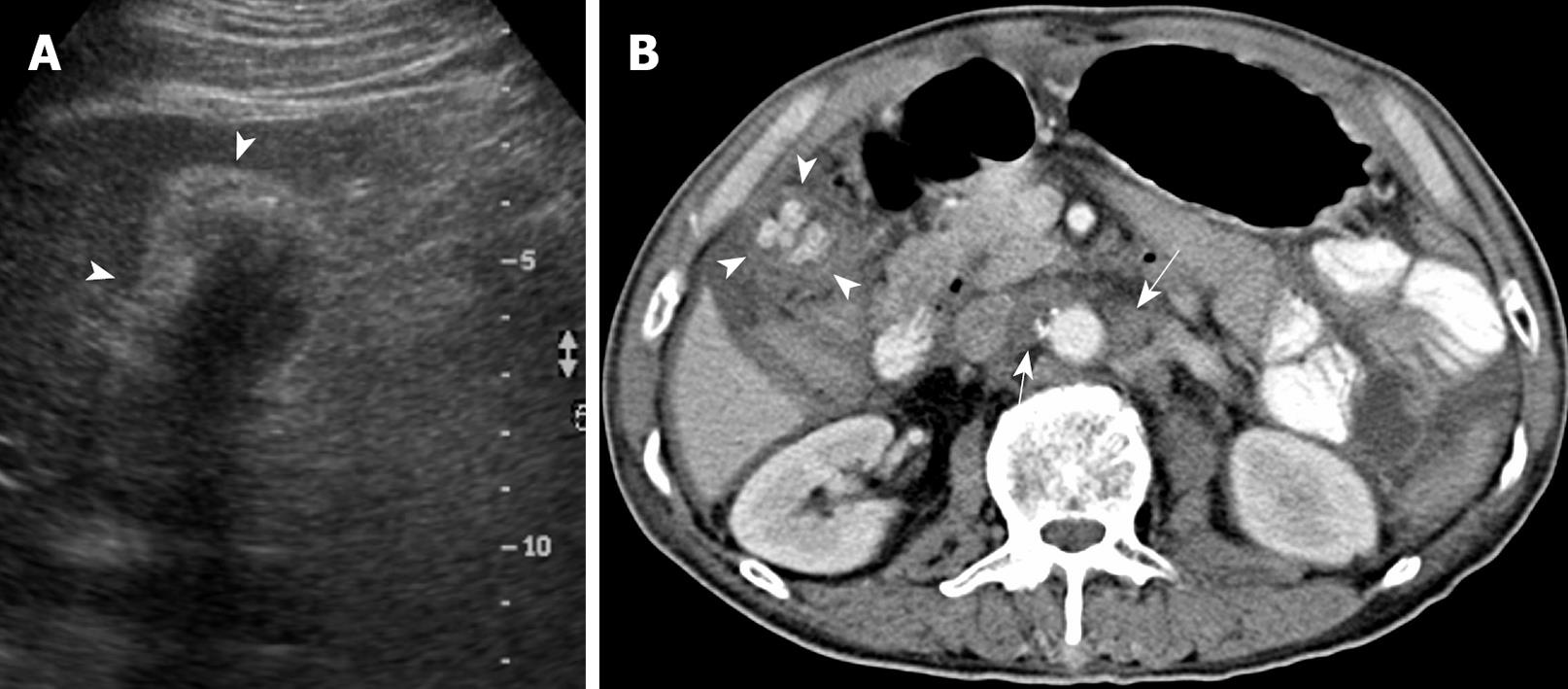
Figure 5 A 78-year-old man admitted to the emergency room with right upper abdominal pain, fever and jaundice for 1 d.
A: US showed a contracted gallbladder with a thickened wall (arrowheads) and multiple gallstones that obliterated underlying details, which mimicked chronic cholecystitis; B: Contrast-enhanced CT showed a relatively small gallbladder with uneven wall thickening (arrowheads), multiple gallstones and pericholecystic inflammatory stranding and fluid suggestive of gallbladder carcinoma (GBCA), with coexistent acute cholecystitis. Note the intercavoaortic and left para-aortic lymph node metastasis (white arrows).
-
Citation: Lee TY, Ko SF, Huang CC, Ng SH, Liang JL, Huang HY, Chen MC, Sheen-Chen SM. Intraluminal
versus infiltrating gallbladder carcinoma: Clinical presentation, ultrasound and computed tomography. World J Gastroenterol 2009; 15(45): 5662-5668 - URL: https://www.wjgnet.com/1007-9327/full/v15/i45/5662.htm
- DOI: https://dx.doi.org/10.3748/wjg.15.5662


