Copyright
©2009 The WJG Press and Baishideng.
World J Gastroenterol. Nov 21, 2009; 15(43): 5493-5497
Published online Nov 21, 2009. doi: 10.3748/wjg.15.5493
Published online Nov 21, 2009. doi: 10.3748/wjg.15.5493
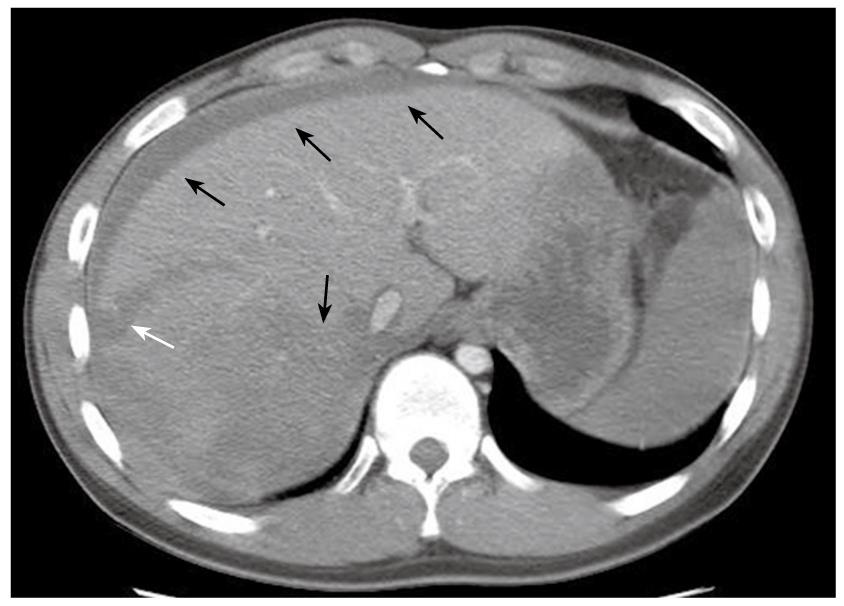
Figure 1 CT of the abdomen showing laceration of the liver right lobe inferior portion (white arrow), with hyperdense fluid in the peritoneal cavity and liver parenchyma, suggesting acute blood collection in these areas (black arrows).
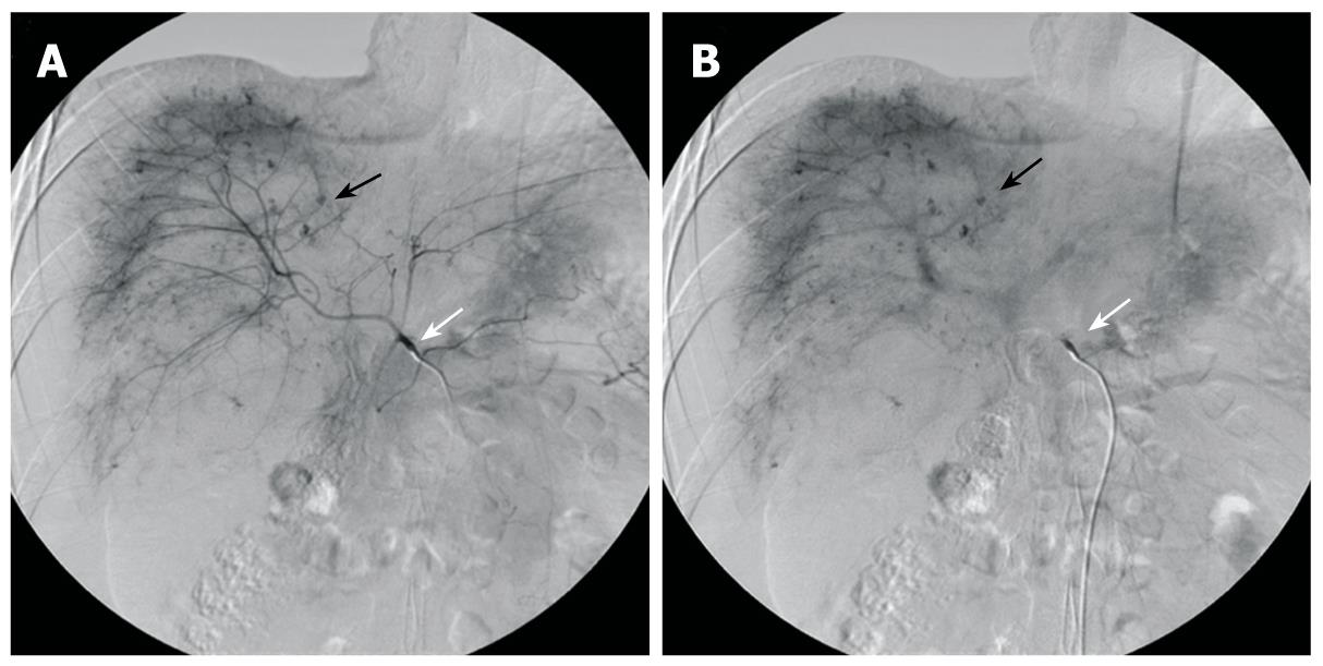
Figure 2 Preoperative selective hepatic angiography.
A: Selective angiography of the celiac trunk (white arrow) shows hemorrhagic spots (black arrow) in hepatic parenchyma at the arterial phase Note: Lesions are mainly distributed in the right hemiliver; B: Angiogram at the portal venous phase shows the lesions with delayed wash-out (black arrow).
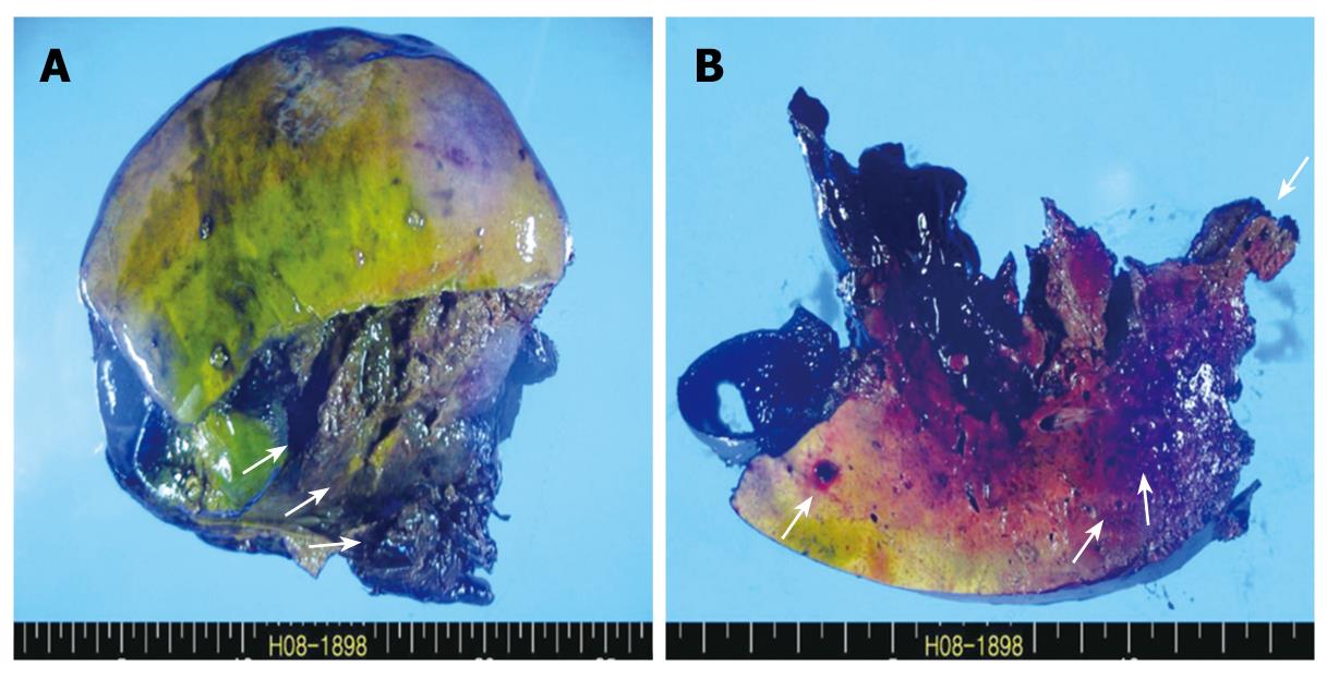
Figure 3 Gross findings.
A: Resected specimen shows extensive necrosis and hemorrhage (arrows); B: Cut surface of the liver parenchyma reveals small blood-filled cystic lesions (arrows).
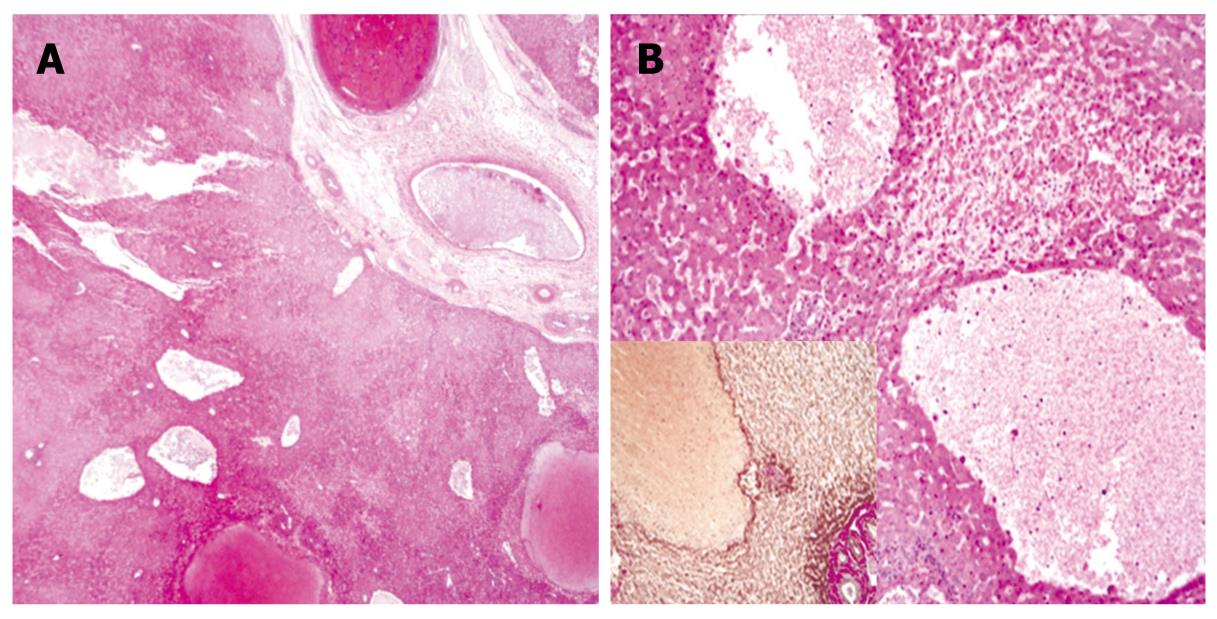
Figure 4 Microscopic findings.
A: Low magnification view shows variable-size, blood-filled cystic spaces (HE, × 10); B: High magnification view shows hemorrhagic necrosis in areas adjacent to peliotic spaces without lining endothelium (HE, × 100); Inset: immunohistochemical staining for reticulin (× 100).
- Citation: Choi SK, Jin JS, Cho SG, Choi SJ, Kim CS, Choe YM, Lee KY. Spontaneous liver rupture in a patient with peliosis hepatis: A case report. World J Gastroenterol 2009; 15(43): 5493-5497
- URL: https://www.wjgnet.com/1007-9327/full/v15/i43/5493.htm
- DOI: https://dx.doi.org/10.3748/wjg.15.5493


