Copyright
©2009 The WJG Press and Baishideng.
World J Gastroenterol. Oct 21, 2009; 15(39): 4907-4914
Published online Oct 21, 2009. doi: 10.3748/wjg.15.4907
Published online Oct 21, 2009. doi: 10.3748/wjg.15.4907
Figure 1 Design of the study.
At the beginning of the experiment, gerbils were inoculated (i.g.) with H pylori (Grps A-E) or vehicle (Brucella broth; Grp F). Until the 21st week, the animals were given drinking water containing no Grp F (open bars) or 50 μg/mL MNNG (Grp A-E). All groups were then switched to distilled water and given a diet containing no drug (Grps A and F) or Celecoxib 10 mg/kg per day for 30 (Grp B), 20 (Grp C), 20 (Grp D) or 15 (Grp E) weeks. The gerbils were sacrificed at week 51.
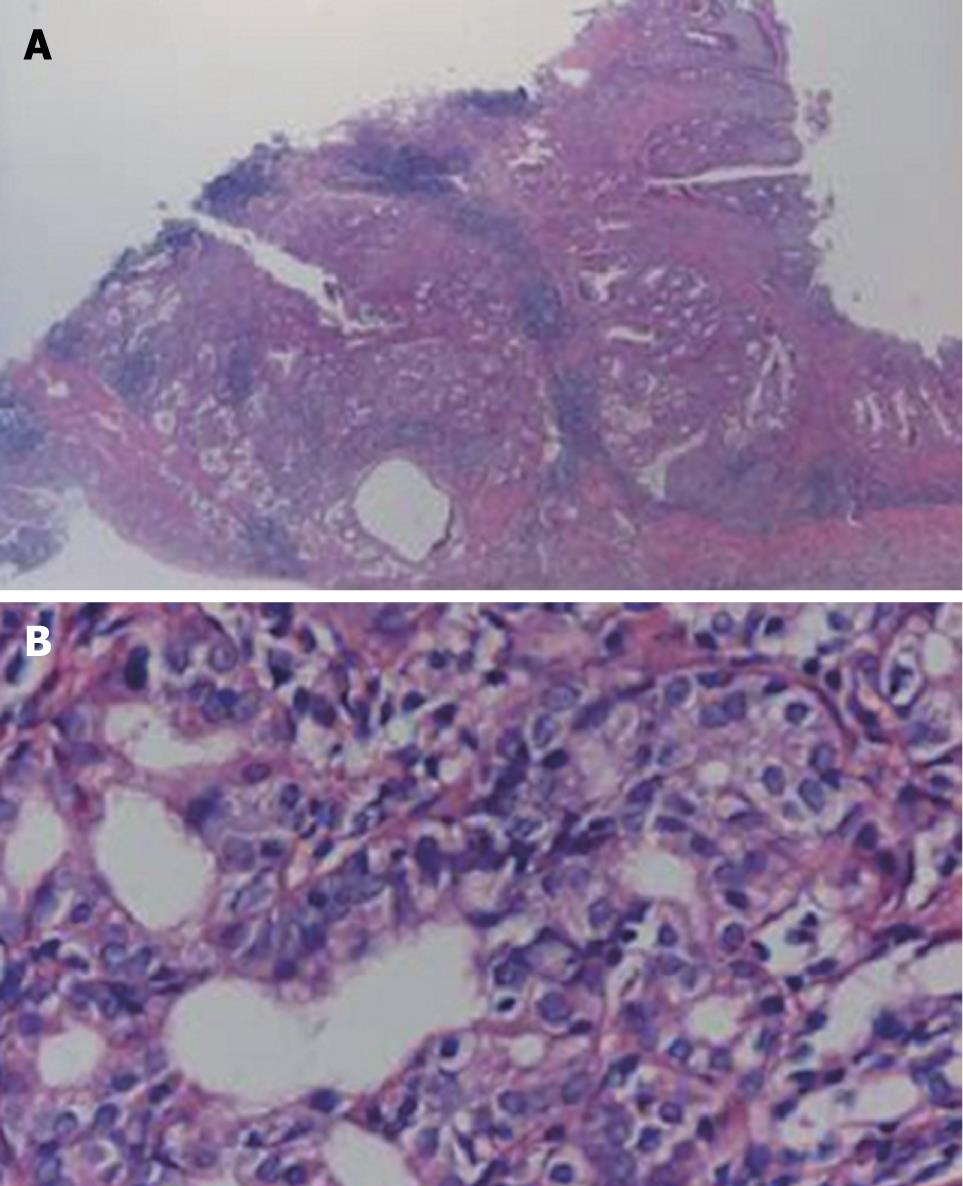
Figure 2 Typical adenocarcinoma in the pyloric mucosa of H pylori-infected MGs.
Shown is a typical well-differentiated adenocarcinoma (A, B) stained with HE. Images were obtained at × 100 (A) and × 400 (B).
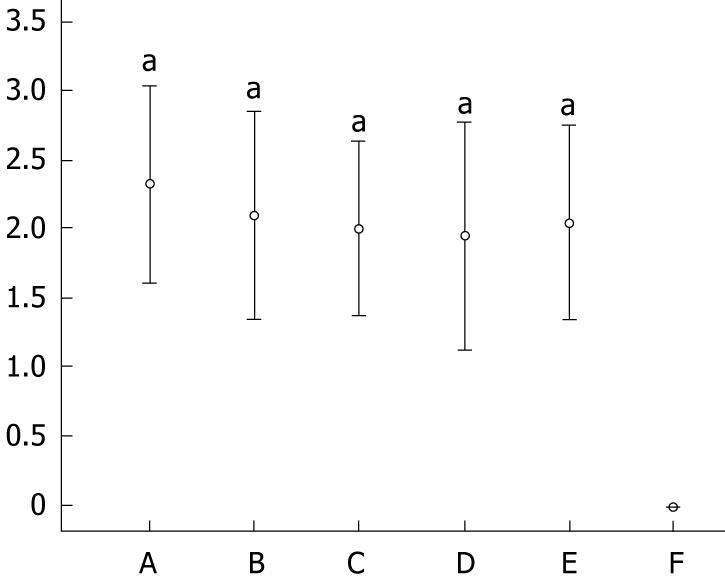
Figure 3 Effect of Celecoxib on inflammation of the stomach mucosa.
No obvious inflammatory change was found in group F. Significantly obvious inflammation was shown in H pylori-infected groups (A-E) vs group F (aP < 0.05), but there was no significant difference among H pylori-infected groups. Group B showed higher inflammatory response than group A, C, D, E.
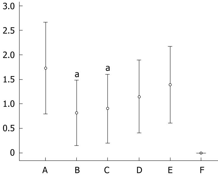
Figure 4 Effect of Celecoxib on the development of intestinal metaplasia (IM).
Severe IM was found in group A. There was significantly lower rates of IM in groups B and C. A relatively lower rate of IM was found in groups D and E (P > 0.05 vs group A). There was no definite IM found in group F. aP < 0.05 vs group A.
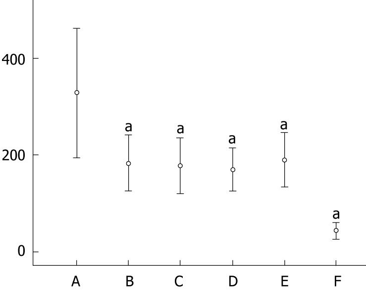
Figure 5 Effect of Celecoxib on the proliferation of gastric mucosal cells in gerbils.
High PCNA positive ratio was found in group A, and the value decreased significantly after treatment with Celecoxib. The value was also lower in group F without H pylori infection. aP < 0.05 vs groups A.
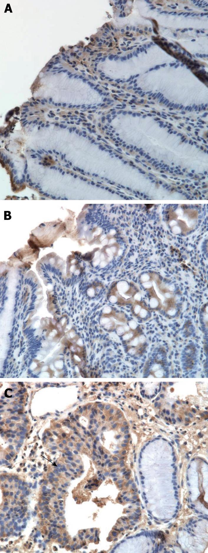
Figure 6 Expression of PCNA by immunohistochemistry method.
A: Normal gastric mucosa; B: Intestinal metaplasia; C: Adenocarcinoma. Adenocarcinoma is pointed out by arrow. Images were obtained at × 400.
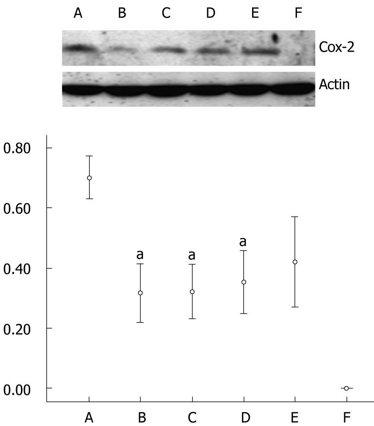
Figure 7 Analysis of COX-2 protein expression in gastric mucosa of gerbils.
The expression of COX-2 protein in group A was high. There was no detectable expression in group F. The expression of COX-2 was inhibited significantly by Celecoxib. There was a significant decrease in groups B, C and D. aP < 0.05 vs group A.
- Citation: Kuo CH, Hu HM, Tsai PY, Wu IC, Yang SF, Chang LL, Wang JY, Jan CM, Wang WM, Wu DC. Short-term Celecoxib intervention is a safe and effective chemopreventive for gastric carcinogenesis based on a Mongolian gerbil model. World J Gastroenterol 2009; 15(39): 4907-4914
- URL: https://www.wjgnet.com/1007-9327/full/v15/i39/4907.htm
- DOI: https://dx.doi.org/10.3748/wjg.15.4907


