Copyright
©2009 The WJG Press and Baishideng.
World J Gastroenterol. Sep 21, 2009; 15(35): 4392-4401
Published online Sep 21, 2009. doi: 10.3748/wjg.15.4392
Published online Sep 21, 2009. doi: 10.3748/wjg.15.4392
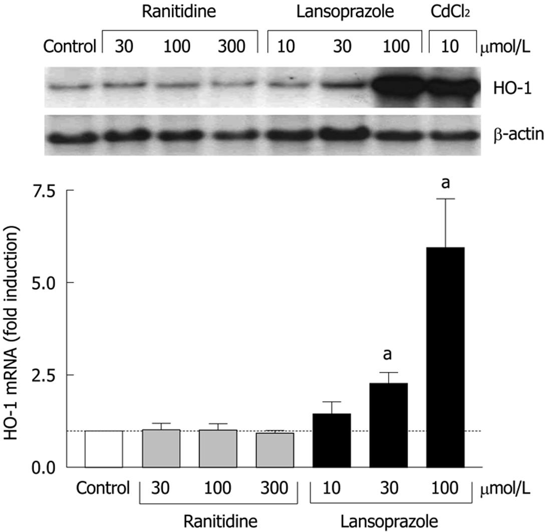
Figure 1 Effect of ranitidine and lansoprazole on HO-1 mRNA induction in endothelial cells after 8 h of incubation.
Fold induction from control levels is shown as mean ± SD of three separate experiments. aP < 0.05 vs control. Treatment with CdCl2 was used as a positive control. Representative Northern blotting analysis is shown in the upper panel.
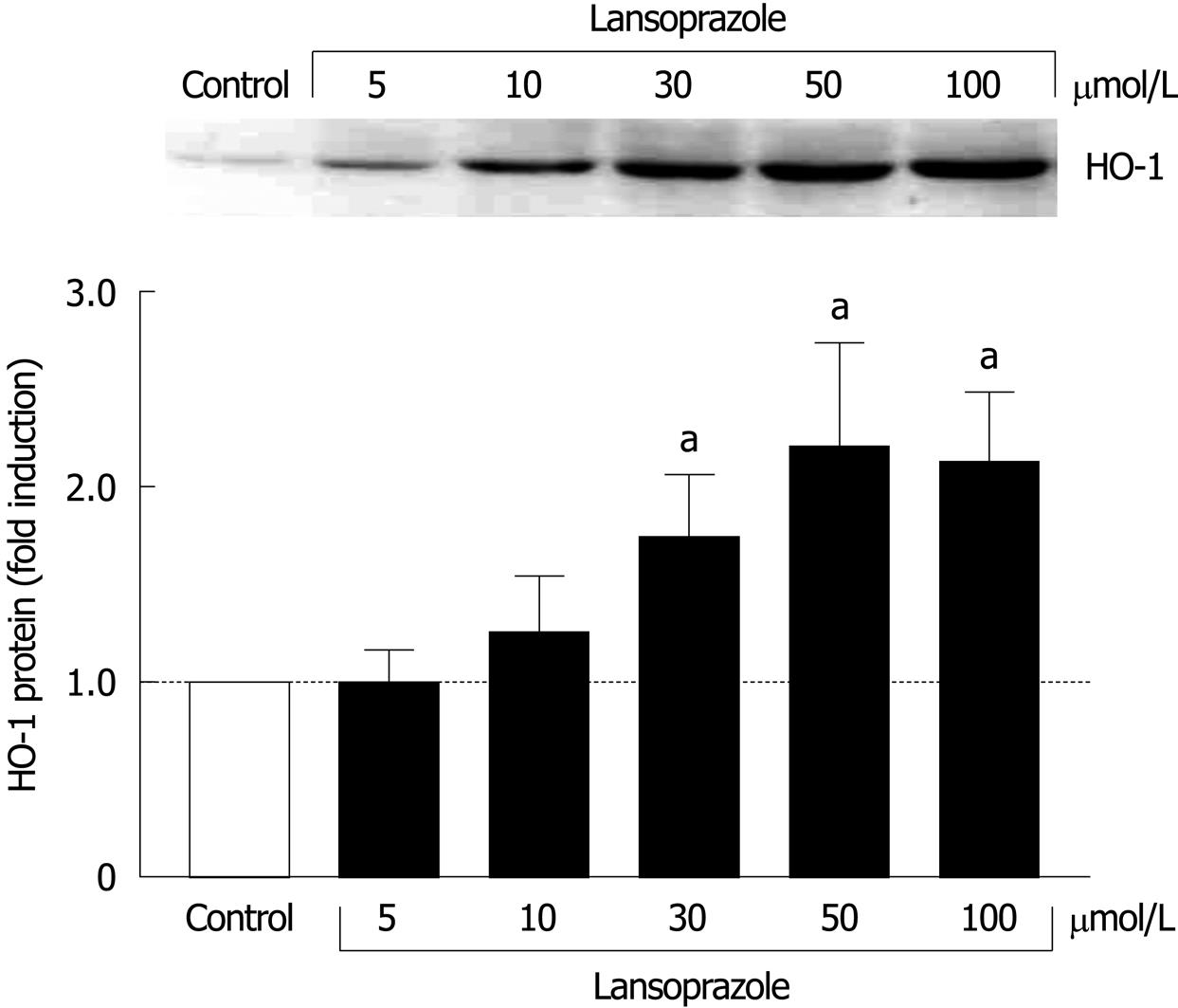
Figure 2 Effect of lansoprazole on HO-1 protein expression in macrophages (J774 cells) after 12 h of incubation.
Fold induction from control levels is shown as mean ± SD of three to six separate experiments. aP < 0.05 vs control. Representative Western blotting analysis is shown in the upper panel.
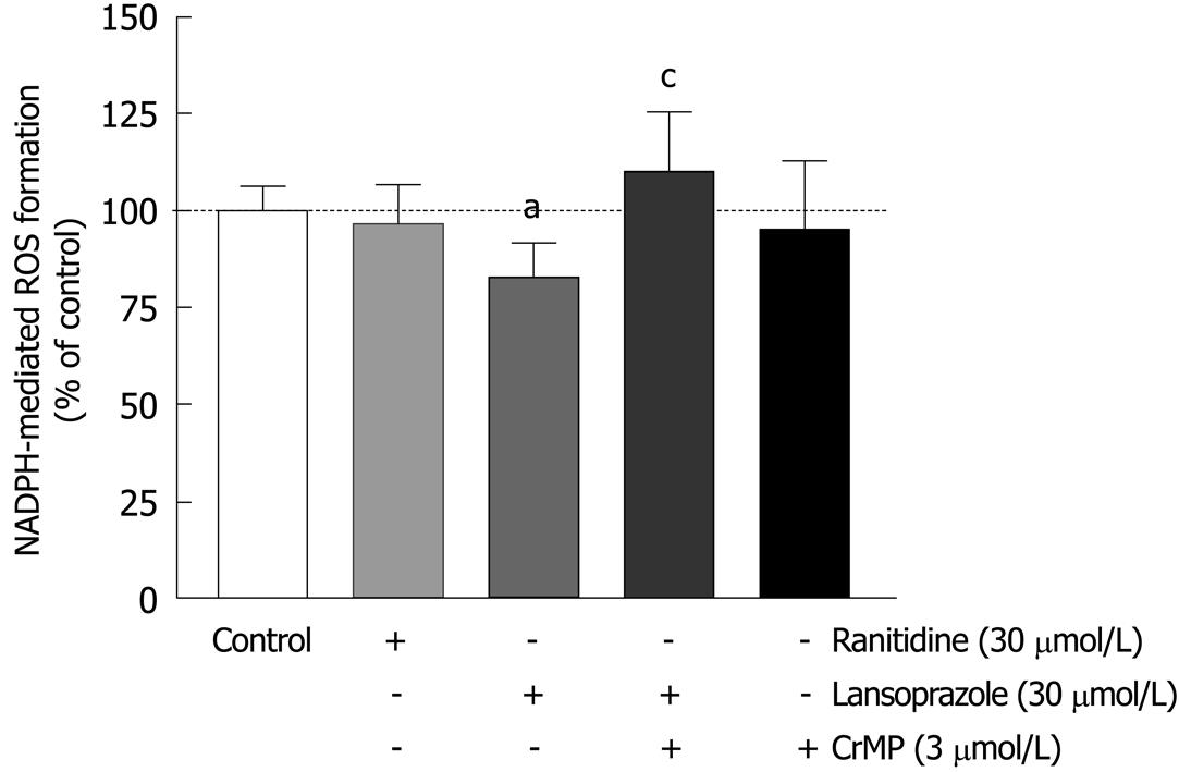
Figure 3 Lansoprazole, but not ranitidine, decreased NADPH-dependent ROS formation in macrophages.
This effect was reversed in the presence of CrMP. Measurements of lucigenin-enhanced chemiluminescence were performed. aP < 0.05 vs control; cP < 0.05 vs lansoprazole alone. Data are shown as mean ± SD of three to six separate experiments.
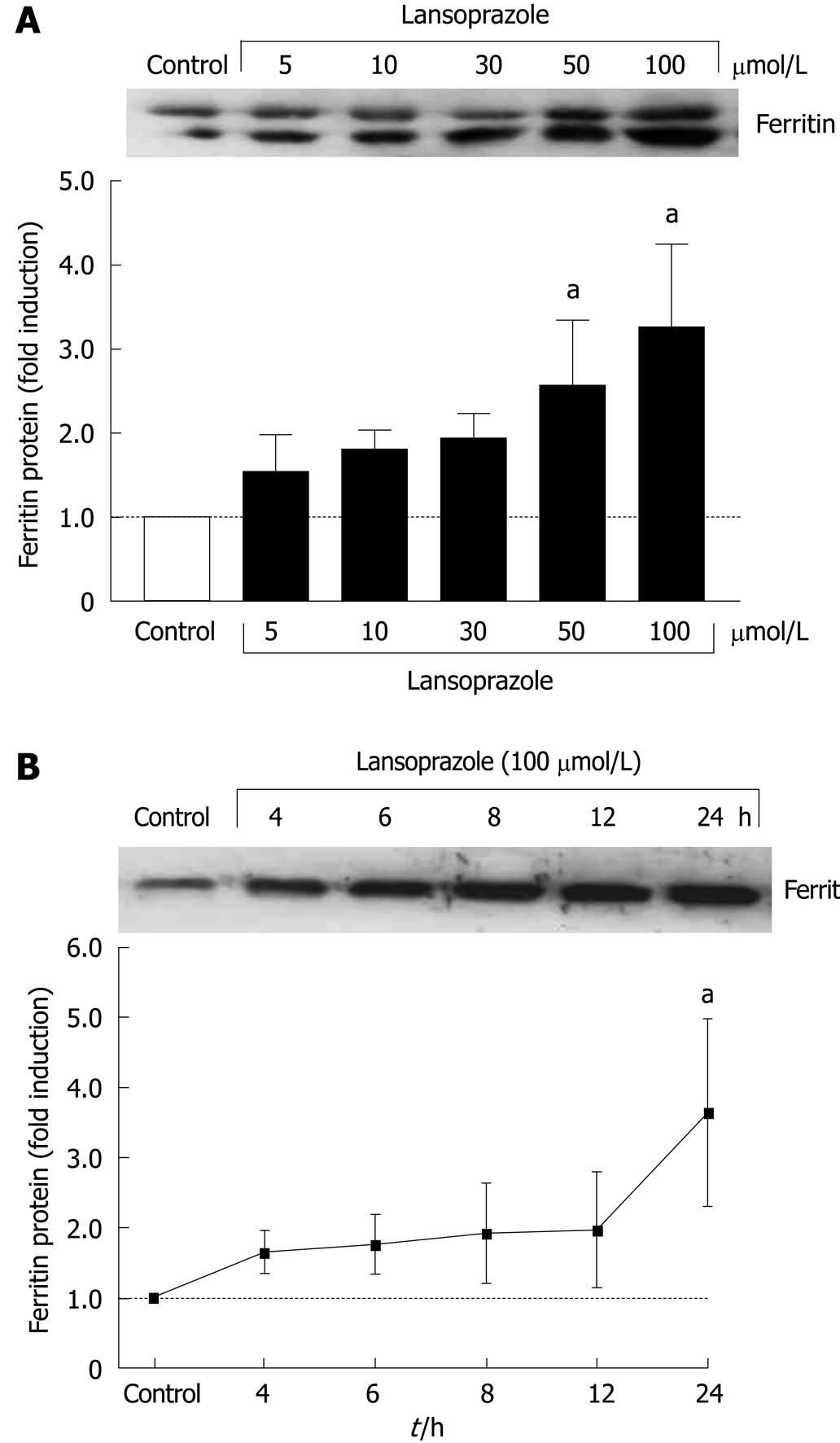
Figure 4 Lansoprazole increased ferritin protein expression in a concentration- and time-dependent manner in macrophages (A) and endothelial cells (B).
Data are shown as mean ± SD of three to six separate experiments. aP < 0.05 vs control. A representative Western blotting analysis is shown in the upper panel.
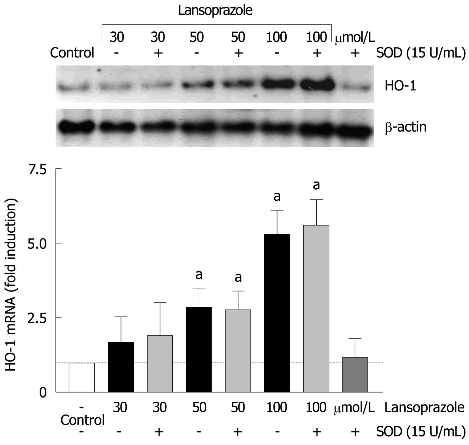
Figure 5 Effect of SOD on lansoprazole-induced HO-1 mRNA expression.
After pretreatment with SOD for 20 min, endothelial cells were incubated with lansoprazole for 8 h. Fold induction compared to control is shown as mean ± SD of three separate experiments. aP < 0.05 vs control. A representative Northern blotting analysis is shown in the upper panel.
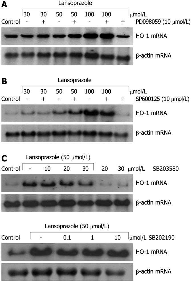
Figure 6 Effect of different MAPK inhibitors on lansoprazole-induced HO-1 mRNA expression.
After pretreatment with ERK inhibitor PD098059 (A), JNK inhibitor SP600125 (B), or p38 inhibitors SB203580 and SB202190 (C) for 20 min, endothelial cells were incubated with lansoprazole for 8 h. The Northern blotting analysis shown is representative of three to six independent experiments.
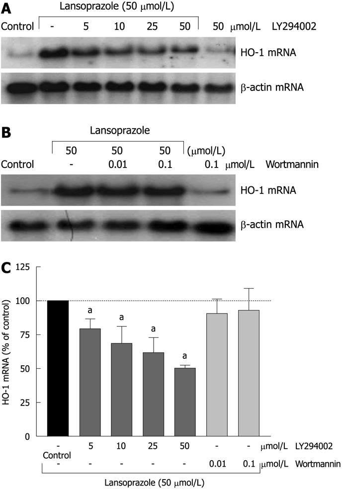
Figure 7 Effect of PI3K inhibitors LY294002 and wortmannin on lansoprazole-induced HO-1 mRNA expression.
After pretreatment with PI3K inhibitors for 20 min, endothelial cells were incubated with lansoprazole for 8 h. Representative Northern blotting analysis is shown (A and B). Results are expressed as mean ± SD of three to six separate experiments compared to lansoprazole (control = gene expression of cells stimulated with 50 μmol/L lansoprazole = 100%) (C). aP < 0.05 vs lansoprazole alone.
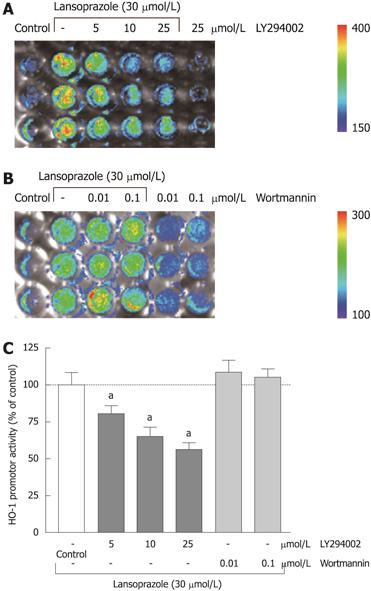
Figure 8 Effect of PI3K-inhibitors LY294002 and wortmannin on lansoprazole-induced HO-1 promoter activity in stable transfected cell lines.
Representative images of three to six separate experiments are shown (A and B). Light emission (shown by the rainbow bars: blue, least intense; red, most intense) was measured in NIH3T3-HO-1-luc cells with the IVIS. Results are expressed as mean ± SD of three to six separate experiments compared to lansoprazole (control = HO-1 promoter activity of cells stimulated with 30 μmol/L lansoprazole = 100%) (C). aP < 0.05 vs lansoprazole alone.
- Citation: Schulz-Geske S, Erdmann K, Wong RJ, Stevenson DK, Schröder H, Grosser N. Molecular mechanism and functional consequences of lansoprazole-mediated heme oxygenase-1 induction. World J Gastroenterol 2009; 15(35): 4392-4401
- URL: https://www.wjgnet.com/1007-9327/full/v15/i35/4392.htm
- DOI: https://dx.doi.org/10.3748/wjg.15.4392


