Copyright
©2009 The WJG Press and Baishideng.
World J Gastroenterol. Sep 14, 2009; 15(34): 4334-4338
Published online Sep 14, 2009. doi: 10.3748/wjg.15.4334
Published online Sep 14, 2009. doi: 10.3748/wjg.15.4334
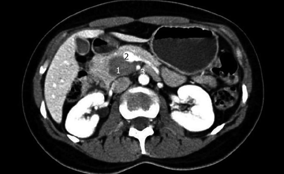
Figure 1 A hypodense and non-infiltrative tumor is occupying the uncinate process of the pancreas (1); It is in close contact with the superior mesenteric vein (2) and superior mesenteric artery (3).
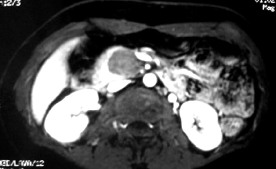
Figure 2 Hypointense-T1 and heterogeneous tumor that does not present any modifications following IV contrast administration with Gadolinium.
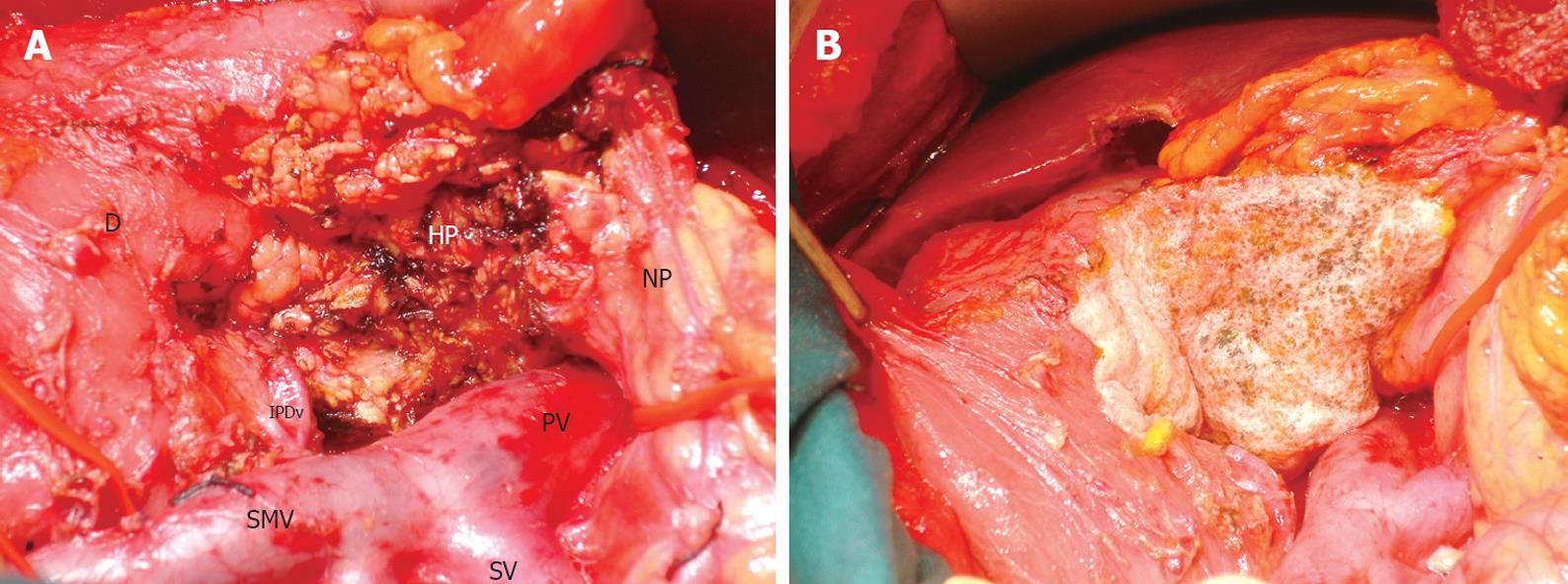
Figure 3 Appearance of surgical field with specimen resected.
A: Uncinate process resected. Duodenum (D), neck of the pancreas (NP) and rest of the head of the pancreas (HP) are preserved. Superior mesenteric vein (SMV) is separated from uncinate process that is removed in the picture. Portal (PV) and splenic vein (SV) are also dissected. Inferior pancreatico-duodenal vessels (IPDv) are preserved; B: TachoSil® sealant is applied over the surface of the surgical bed for prevention of pancreatic fistula.
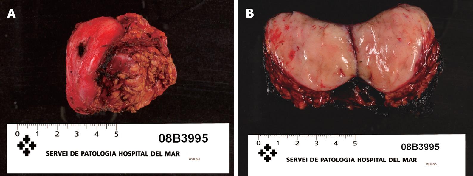
Figure 4 Macroscopic appearance of the surgical specimen.
A: Nodular lesion with well-demarcated margins with normal residual pancreatic tissue adjoining; B: The tumor is well-circumscribed and firm. The inner surface is tan without evidence of necrosis or hemorrhage.
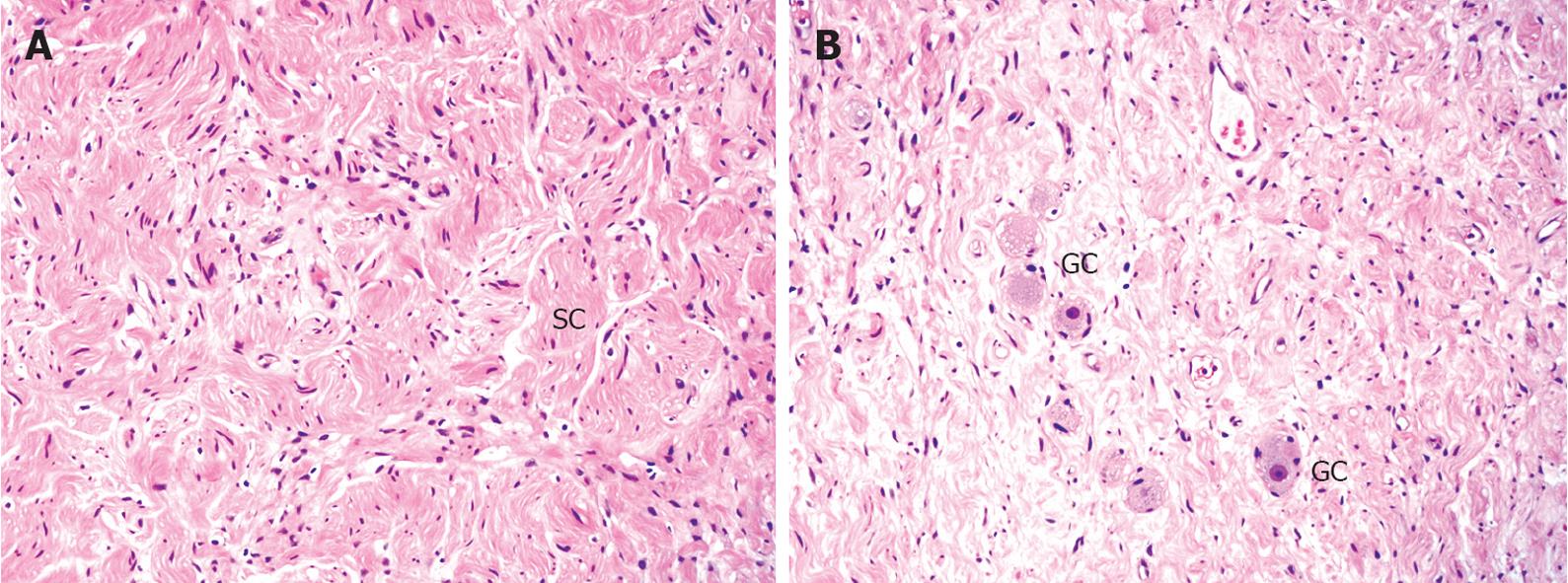
Figure 5 Microscopic images of the tumor (HE, × 10).
A: The lesion is composed of a proliferation of spindle-shaped cells in a whorled or fascicular pattern [Schwann cells (SC) and nerve fibers]. The cells have elongated and wavy nuclei with eosinophilic cytoplasm; B: Scattered mature ganglion cells (GC) are another of the histopathologic components of the tumor.
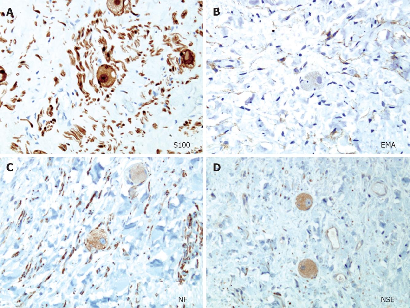
Figure 6 Representative immunocytochemistry samples of the typical ganglioneuroma (× 20).
A: S100 protein stain showing mostly Schwann cells and ganglion cells; B, C: Epithelial membrane antigen (EMA) and neurofilament (NF) stain showing the nerve fibers, highlighting the perineural cells and the axons of the nerve fibers; D: Ganglion cells are positive for neuronal-specific enolase (NSE).
- Citation: Poves I, Burdío F, Iglesias M, Martínez-Serrano MLÁ, Aguilar G, Grande L. Resection of the uncinate process of the pancreas due to a ganglioneuroma. World J Gastroenterol 2009; 15(34): 4334-4338
- URL: https://www.wjgnet.com/1007-9327/full/v15/i34/4334.htm
- DOI: https://dx.doi.org/10.3748/wjg.15.4334


