Copyright
©2009 The WJG Press and Baishideng.
World J Gastroenterol. Sep 7, 2009; 15(33): 4215-4217
Published online Sep 7, 2009. doi: 10.3748/wjg.15.4215
Published online Sep 7, 2009. doi: 10.3748/wjg.15.4215
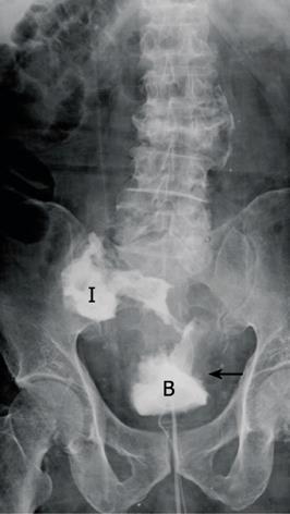
Figure 1 Cystography showed an enterovesical fistula (arrow).
B: Urinary bladder; I: Ileum.
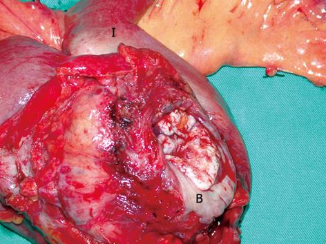
Figure 2 Surgical specimen of urinary bladder dome and ileum resected en bloc.
B: Urinary bladder; I: Ileum.
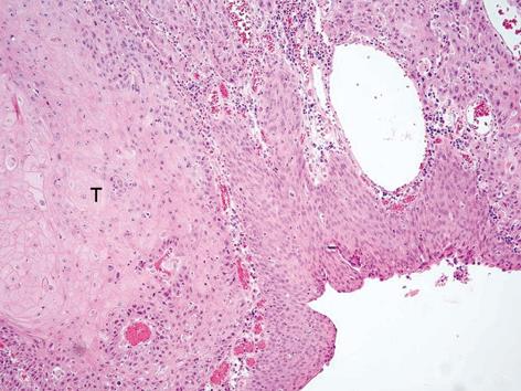
Figure 3 Microscopic examination yielded the diagnosis of squamous cell carcinoma of the urinary bladder.
HE staining, original magnification × 40. T: Tumor burden.
- Citation: Ou Yang CH, Liu KH, Chen TC, Chang PL, Yeh TS. Enterovesical fistula caused by a bladder squamous cell carcinoma. World J Gastroenterol 2009; 15(33): 4215-4217
- URL: https://www.wjgnet.com/1007-9327/full/v15/i33/4215.htm
- DOI: https://dx.doi.org/10.3748/wjg.15.4215


