Copyright
©2009 The WJG Press and Baishideng.
World J Gastroenterol. Sep 7, 2009; 15(33): 4199-4200
Published online Sep 7, 2009. doi: 10.3748/wjg.15.4199
Published online Sep 7, 2009. doi: 10.3748/wjg.15.4199
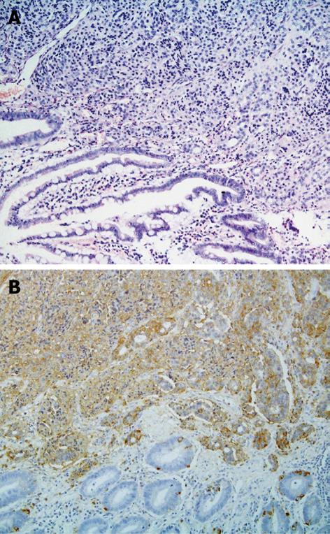
Figure 1 Histopathological examination.
A: Mixed carcinoid-adenocarcinoma neoplasm (× 200); B: Chromogranin A positive stain of tumour cells (× 200).
- Citation: Musialik JA, Kohut MJ, Marek T, Wodołażski A, Hartleb M. Composite neuroendocrine and adenomatous carcinoma of the papilla of Vater. World J Gastroenterol 2009; 15(33): 4199-4200
- URL: https://www.wjgnet.com/1007-9327/full/v15/i33/4199.htm
- DOI: https://dx.doi.org/10.3748/wjg.15.4199


