Copyright
©2009 The WJG Press and Baishideng.
World J Gastroenterol. Sep 7, 2009; 15(33): 4105-4110
Published online Sep 7, 2009. doi: 10.3748/wjg.15.4105
Published online Sep 7, 2009. doi: 10.3748/wjg.15.4105
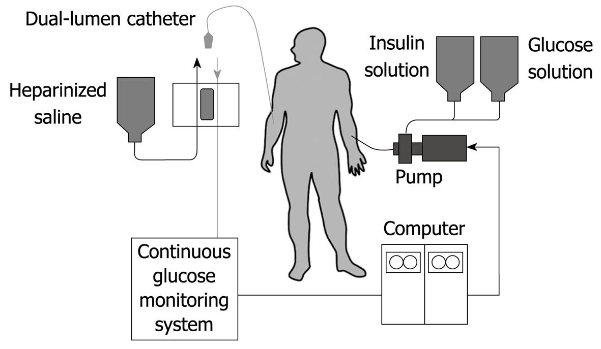
Figure 1 Schematic diagram of the bedside-type artificial endocrine pancreas.
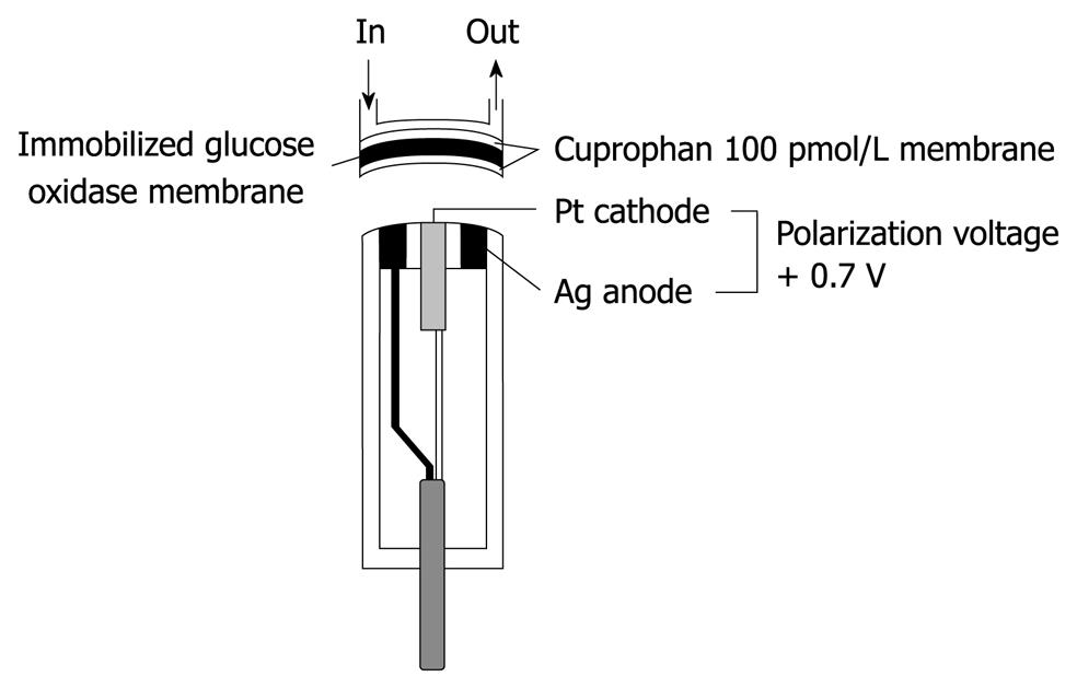
Figure 2 Structure of a glucose sensor of the bedside-type artificial endocrine pancreas.
The immobilized glucose oxidase was covered with hydrophilic Cuprophan 100 pmol/L.
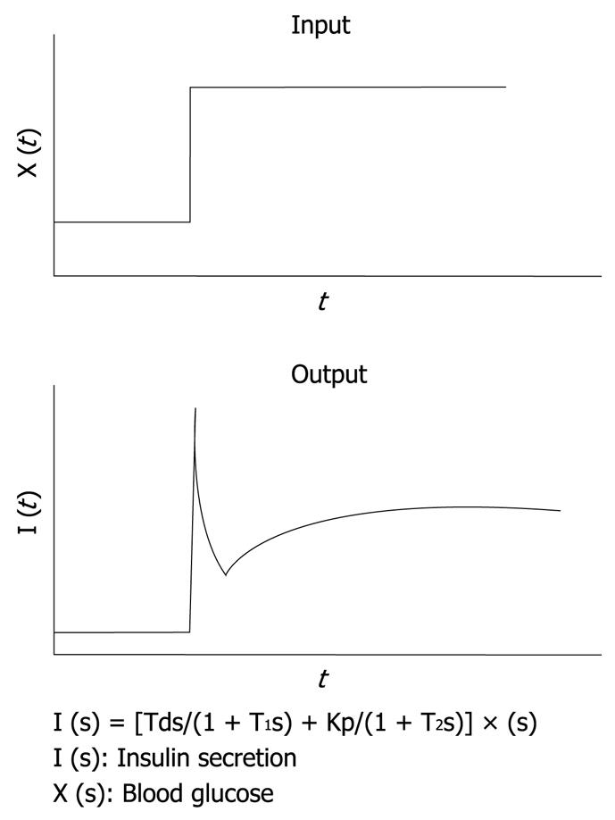
Figure 3 Mathematical model for insulin infusion algorithm.
The stepwise input of glucose concentration [X (t)] and biphasic response of insulin secretion [I (t)] are depicted. The relationship between input and output is expressed in the transfer function. I (s), X (s) and dX (s) are the Laplace transformed form of I (t), X (t) and dX (t)/dt, respectively. T1 and T2 are the first order delay in response.
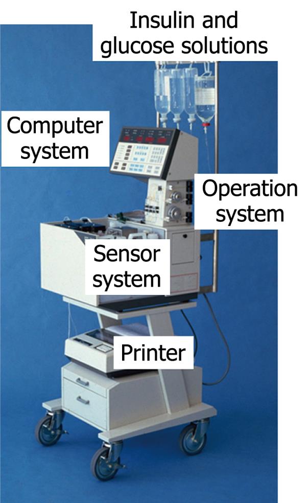
Figure 4 Bedside-type artificial endocrine pancreas (STG-22, Nikkiso Co.
Ltd. Japan).
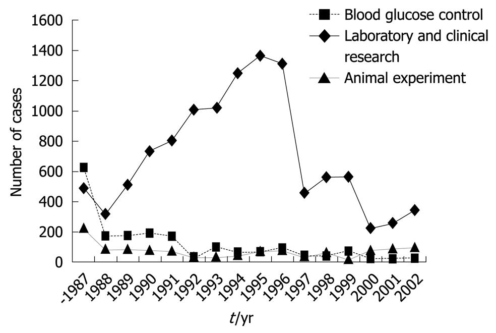
Figure 5 Number of cases to whom the bedside-type artificial endocrine pancreas had been applied in Japan.
Artificial Organs Registry Report in Japan, 2002.
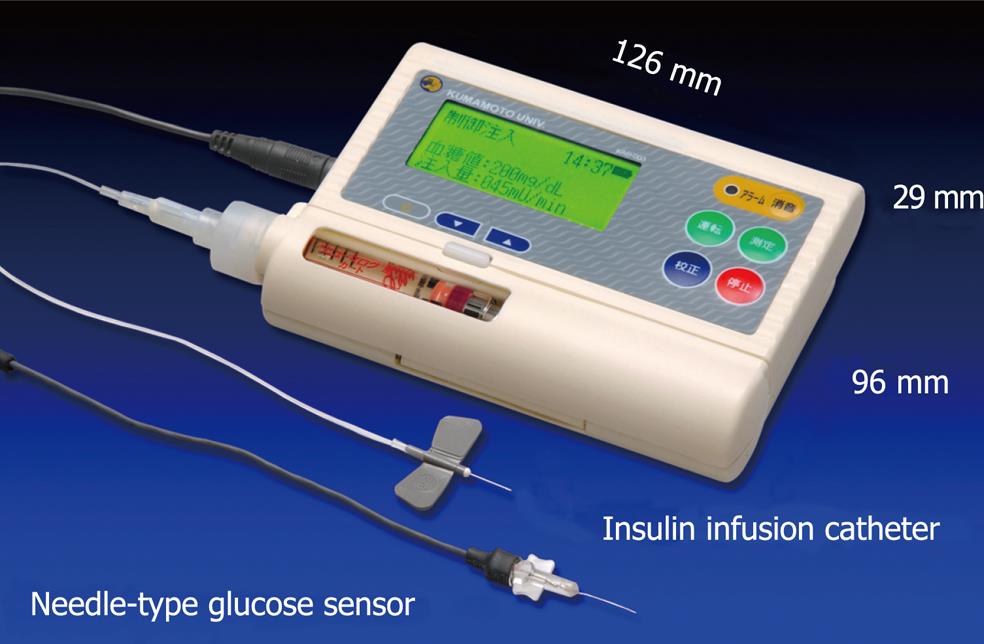
Figure 6 Newly designed wearable artificial endocrine pancreas.
The total system is packed into a small unit (12.6 cm ¡Á 2.9 cm ¡Á 9.6 cm) weighing 250 g (KAP-003, Nikkiso). A glucose sensor is placed in the subcutaneous tissue and measures glucose concentration continuously. The blood glucose concentration (BG, in mg/dL) and the insulin infusion rate (IIR, in mU/min) appear every minute on a large LCD display.
- Citation: Nishida K, Shimoda S, Ichinose K, Araki E, Shichiri M. What is artificial endocrine pancreas? Mechanism and history. World J Gastroenterol 2009; 15(33): 4105-4110
- URL: https://www.wjgnet.com/1007-9327/full/v15/i33/4105.htm
- DOI: https://dx.doi.org/10.3748/wjg.15.4105


