Copyright
©2009 The WJG Press and Baishideng.
World J Gastroenterol. Aug 28, 2009; 15(32): 4083-4086
Published online Aug 28, 2009. doi: 10.3748/wjg.15.4083
Published online Aug 28, 2009. doi: 10.3748/wjg.15.4083
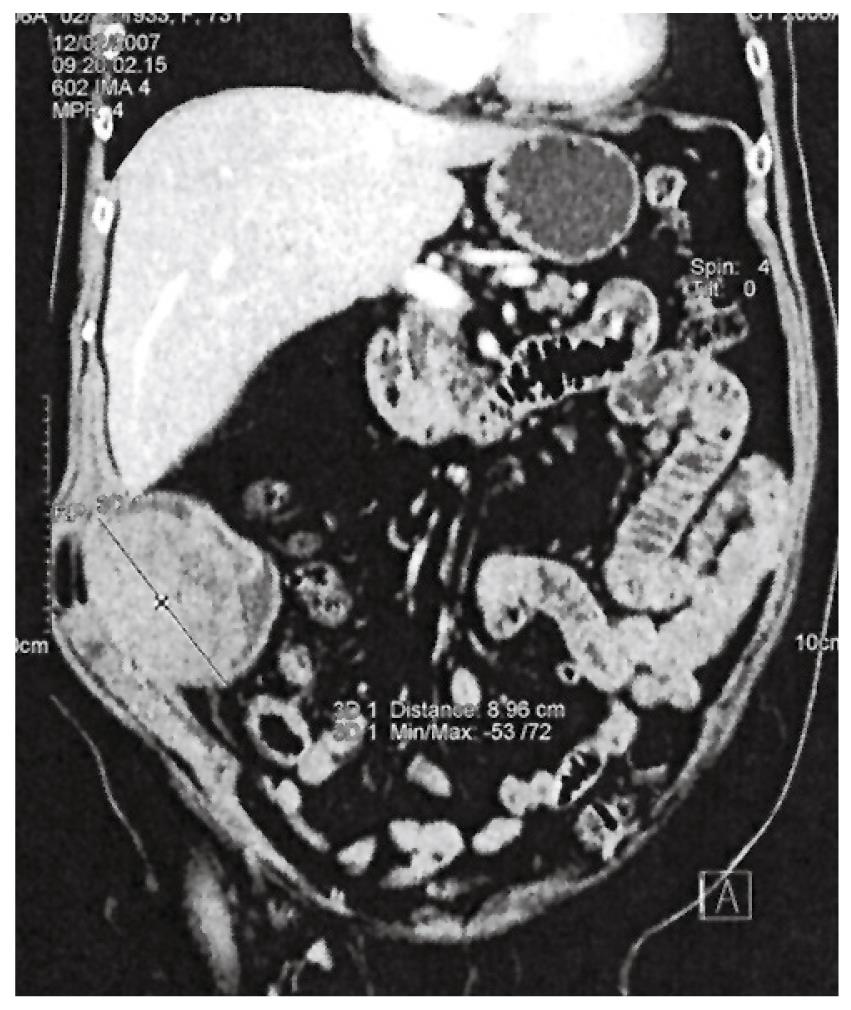
Figure 1 CT scan of the abdominal wall mass in the right lower abdomen protruding into the abdominal cavity dislocating small bowel loops.
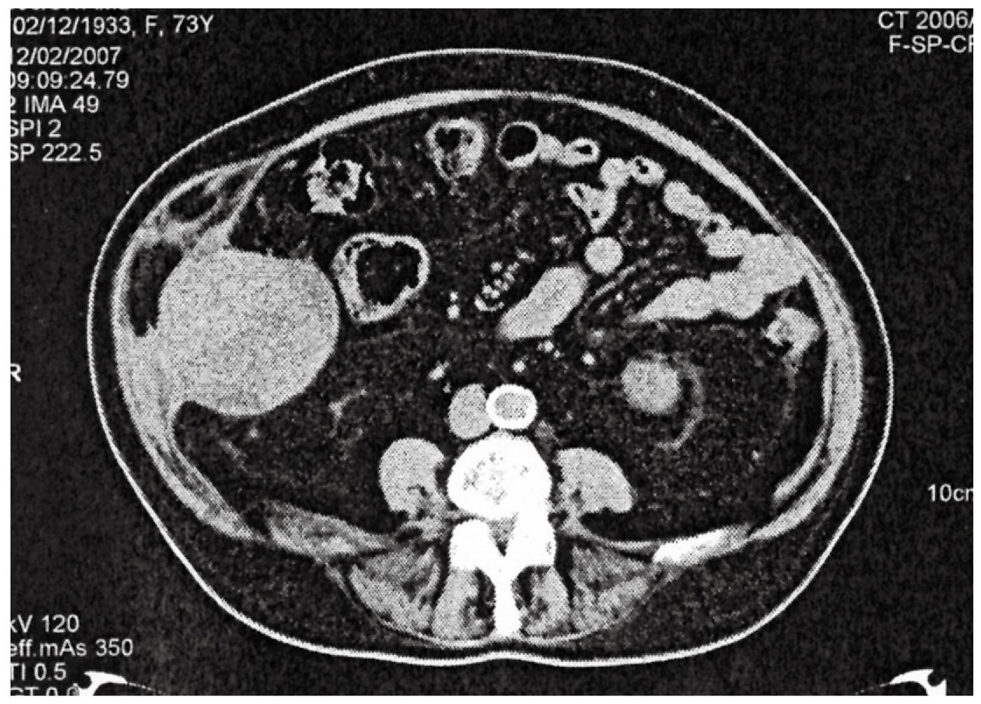
Figure 2 CT scan of the lower abdomen showing the abdominal wall mass with thickened and bulging abdominal wall and no communication with the cecum.
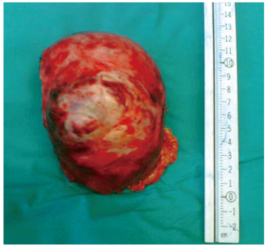
Figure 3 Excised abdominal wall mass.
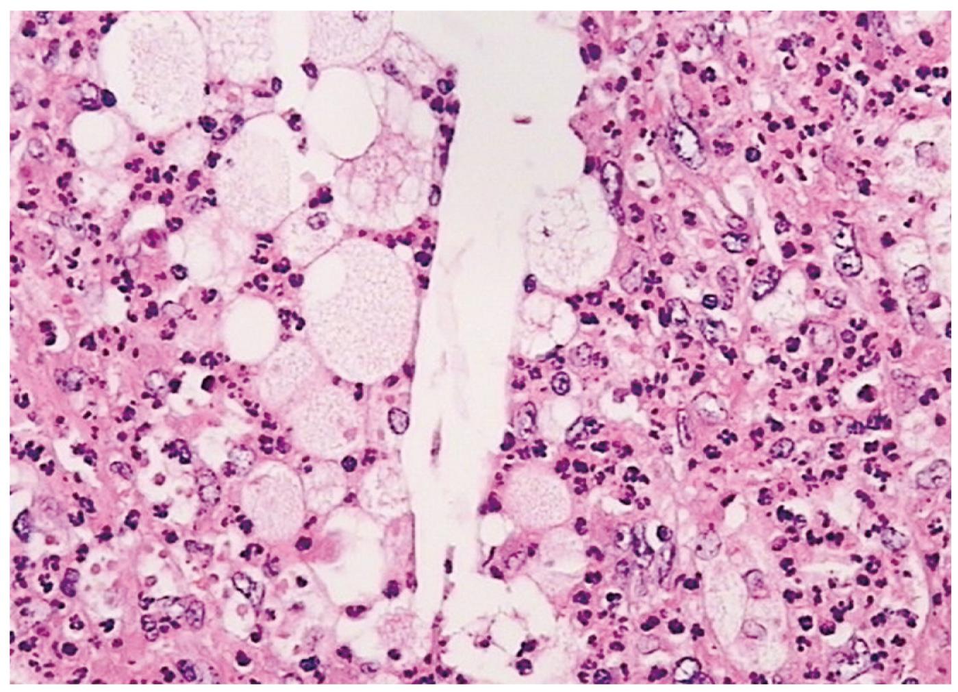
Figure 4 Histological image showing the abscess with polymorphonuclear leukocytes, histiocytes and cholesterol crystals (central part of the photograph).
(HE, × 400).
- Citation: Augustin G, Korolija D, Skegro M, Jakic-Razumovic J. Suture granuloma of the abdominal wall with intra-abdominal extension 12 years after open appendectomy. World J Gastroenterol 2009; 15(32): 4083-4086
- URL: https://www.wjgnet.com/1007-9327/full/v15/i32/4083.htm
- DOI: https://dx.doi.org/10.3748/wjg.15.4083


