Copyright
©2009 The WJG Press and Baishideng.
World J Gastroenterol. Aug 28, 2009; 15(32): 4016-4022
Published online Aug 28, 2009. doi: 10.3748/wjg.15.4016
Published online Aug 28, 2009. doi: 10.3748/wjg.15.4016
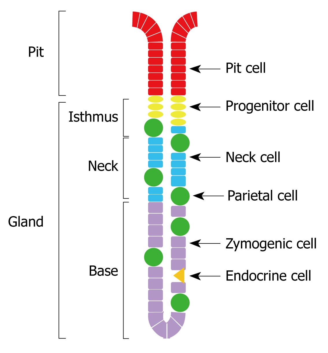
Figure 1 Schematic drawing of the structural unit of the gastric epithelium showing the pit and three glandular regions and the scattered H,K-ATPase-containing parietal cells.
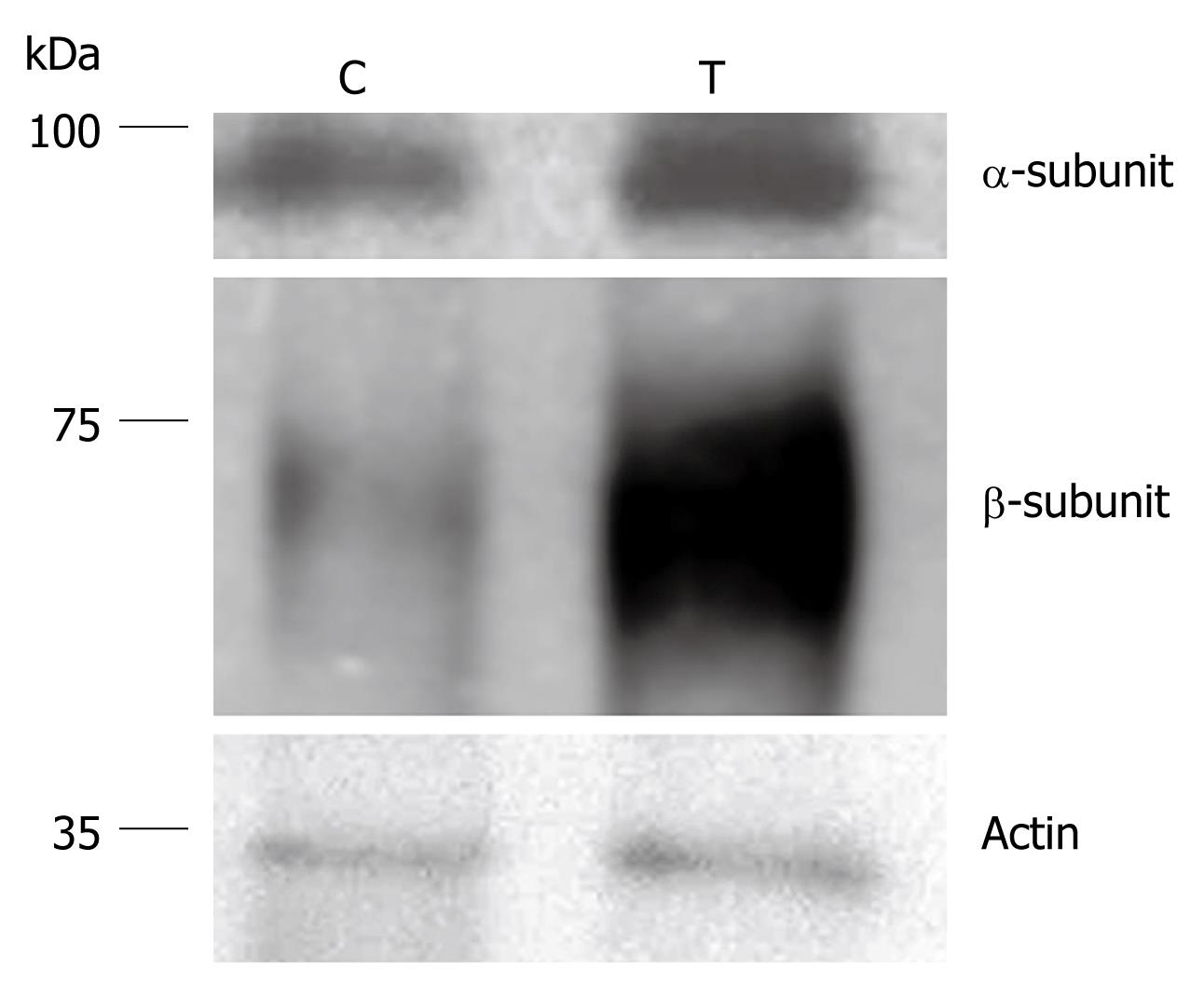
Figure 2 Representative transblots showing protein expression of the α- and β-subunits of H,K-ATPase in control (C) and smoke-treated (T) mice.
Note the differences in the intensity of the protein bands in control vs smoke-treated samples. Ten micrograms of proteins were loaded per lane. Beta actin was detected by a mouse monoclonal antibody and used as a loading control.
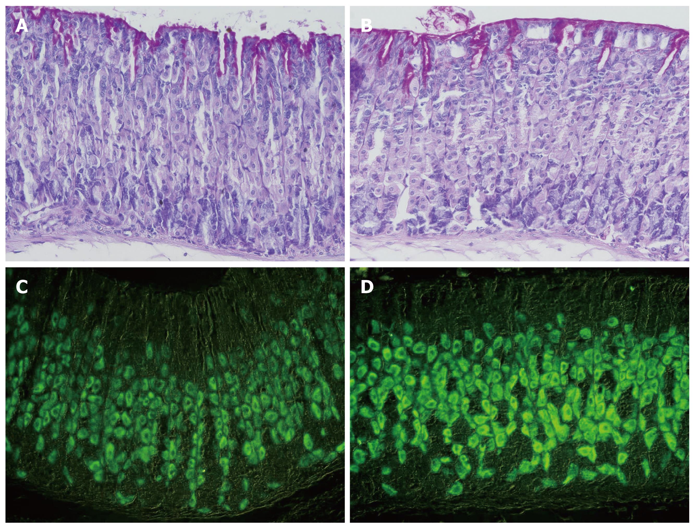
Figure 3 Gastric mucosal tissue sections of control (A, C) and smoke-treated (B, D) mice.
A and B demonstrate the gastric mucosae of control and treated mice stained with periodic acid-Schiff and hematoxylin. No difference is noted in parietal cells of control and treated tissues. Immunohistochemical labeling of parietal cells in the gastric mucosa of control (C) and smoke-treated (D) mice with antibodies specific for H,K-ATPase β-subunit. Labeled parietal cells are distributed throughout the gastric glands. Note the difference in the labeling intensity of parietal cells in control vs smoke-treated mice, × 400.
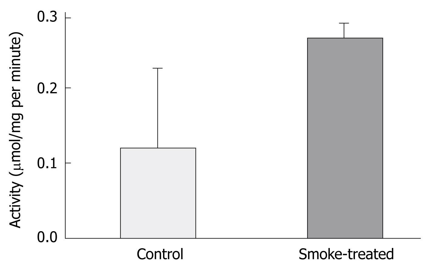
Figure 4 Analysis of the enzymatic activity of the H,K-ATPase of gastric mucosae in control and smoke-treated mice.
Note the increased activity in the cigarette smoke-treated homogenate.
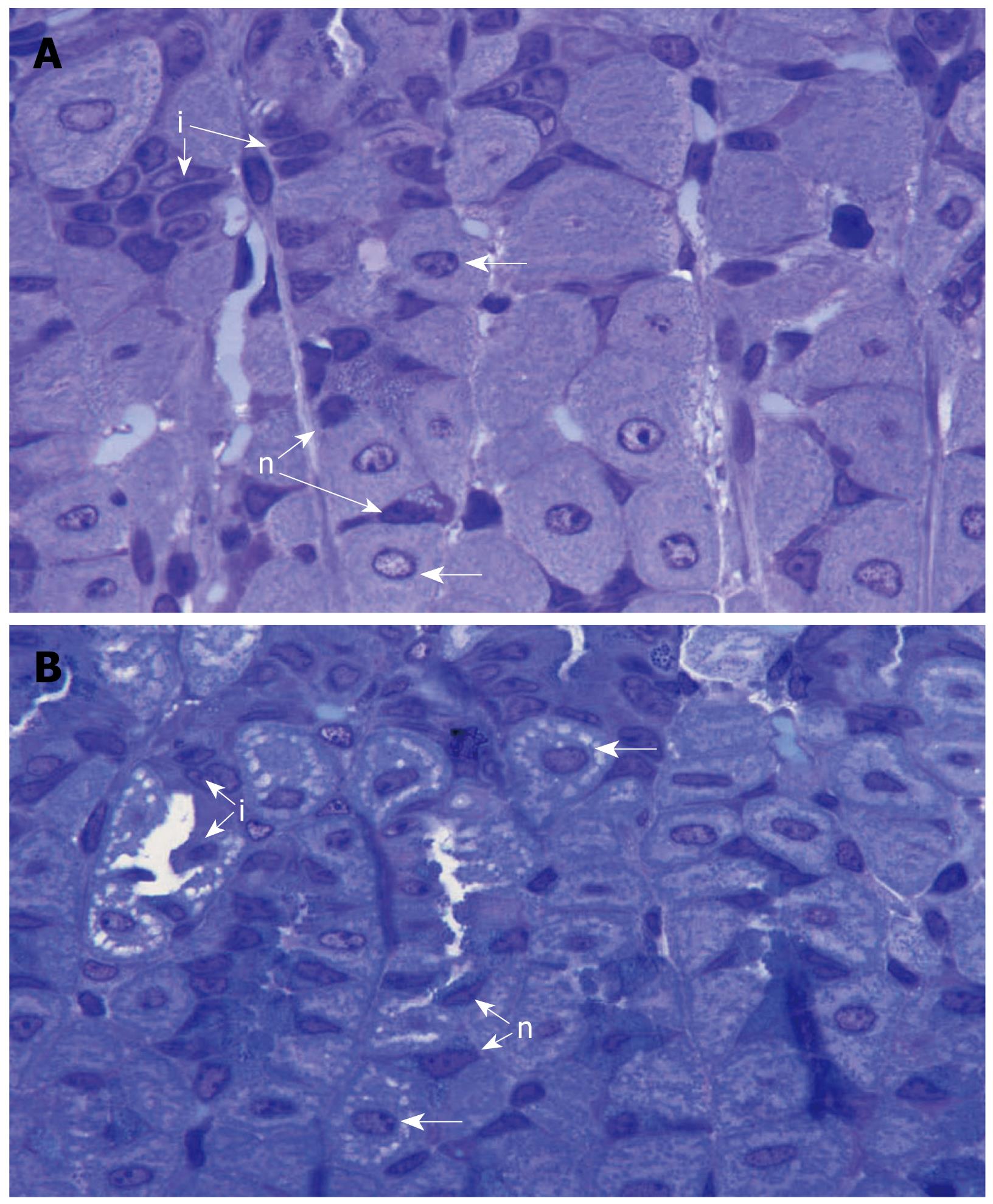
Figure 5 Semithin (0.
5-micron-thick) sections of the gastric mucosae of control (A) and smoke-treated (B) mice stained with toluidine blue to demonstrate the isthmus and neck regions of the gastric glands. The large numerous parietal cells (horizontal arrows) are separated by progenitor or isthmal cells (i) and neck cells (n). Note that, in the smoke-treated tissue but not control tissue, there are pale areas in the cytoplasm of parietal cells which represent expanded lumen of the intracellular canaliculi, × 1000.
- Citation: Hammadi M, Adi M, John R, Khoder GA, Karam SM. Dysregulation of gastric H,K-ATPase by cigarette smoke extract. World J Gastroenterol 2009; 15(32): 4016-4022
- URL: https://www.wjgnet.com/1007-9327/full/v15/i32/4016.htm
- DOI: https://dx.doi.org/10.3748/wjg.15.4016


