Copyright
©2009 The WJG Press and Baishideng.
World J Gastroenterol. Aug 28, 2009; 15(32): 3992-3998
Published online Aug 28, 2009. doi: 10.3748/wjg.15.3992
Published online Aug 28, 2009. doi: 10.3748/wjg.15.3992
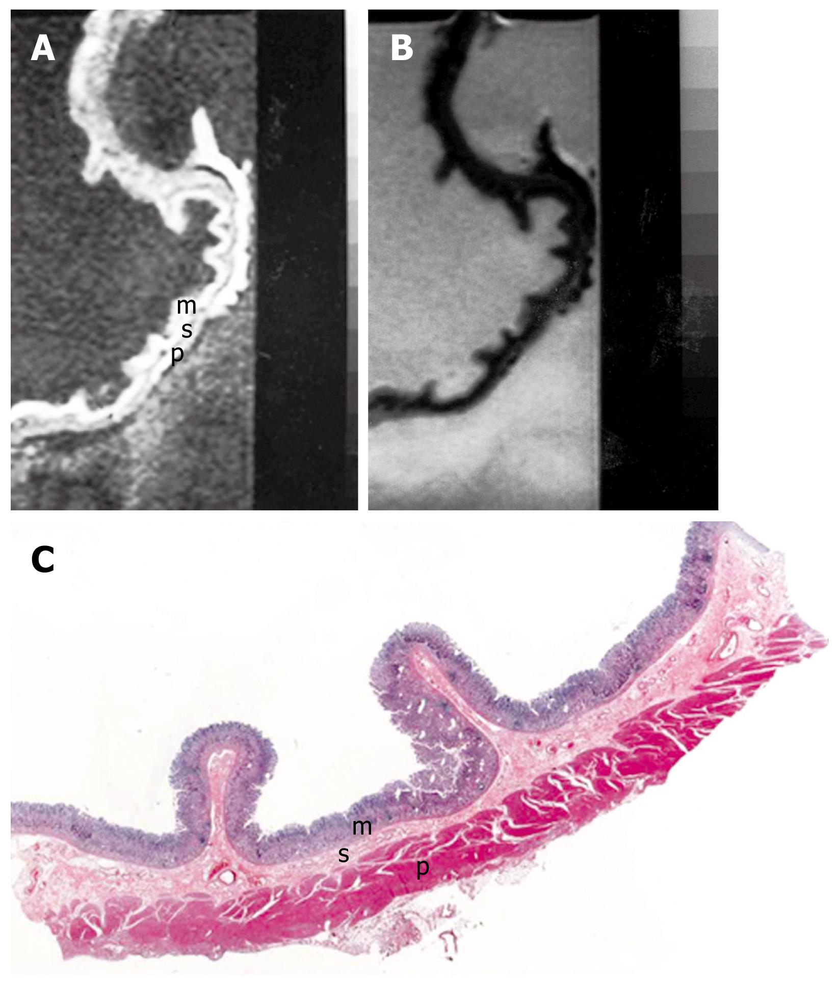
Figure 1 MRI and histology of normal gastric wall.
A: T1-weighted (500/20) sagittal image of resected gastric wall showed three layers. The inner layer corresponds to the mucosa (m) and the middle layer to the submucosa (s). The outer layer basically consists of the muscularis propria (p) from which the serosa cannot be differentiated; B: T2-weighted (2500/90) MRI showed low SI on mucosa and muscularis propria and relatively high SI on submucosa; C: Light microscopic section of normal gastric wall obtained from the greater curvature site of stomach body showed three layers which are compatible with inner mucosal layer (m), middle submucosa layer (s) and outer muscularis propria and serosal layer (p) (HE stain; original magnification, × 1).
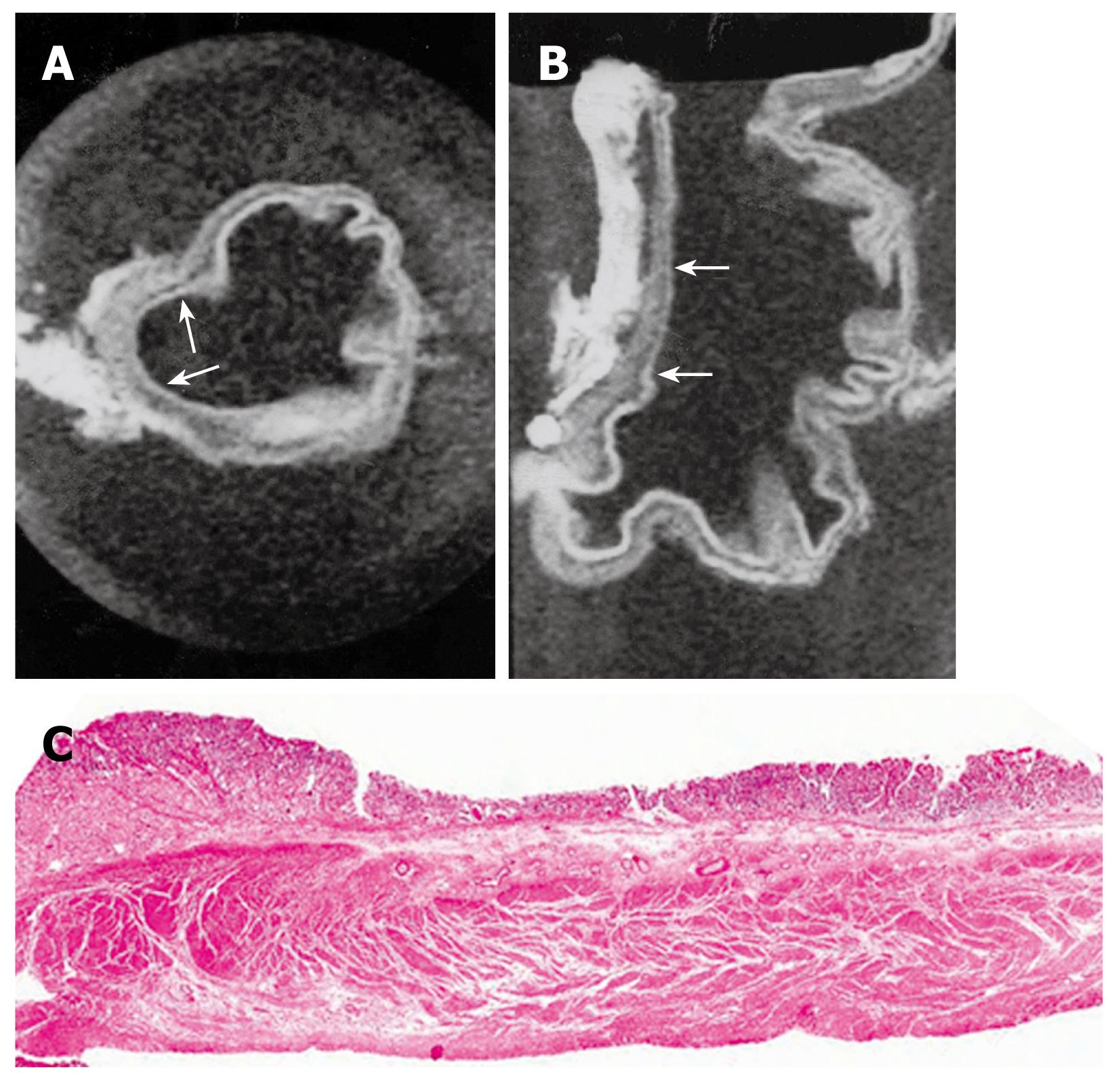
Figure 2 MRI and histology of early gastric cancer.
A: T1-weighted (500/20) axial image showed depression of gastric wall and obliteration of submucosal low SI (arrows); B: T1-weighted sagittal MRI showed depressed mucosa with tumor invasion to submucosa layer (arrows); C: Light microscopic section showed depressed mucosa with tumor invasion to submucosa (HE stain; original magnification, × 1).
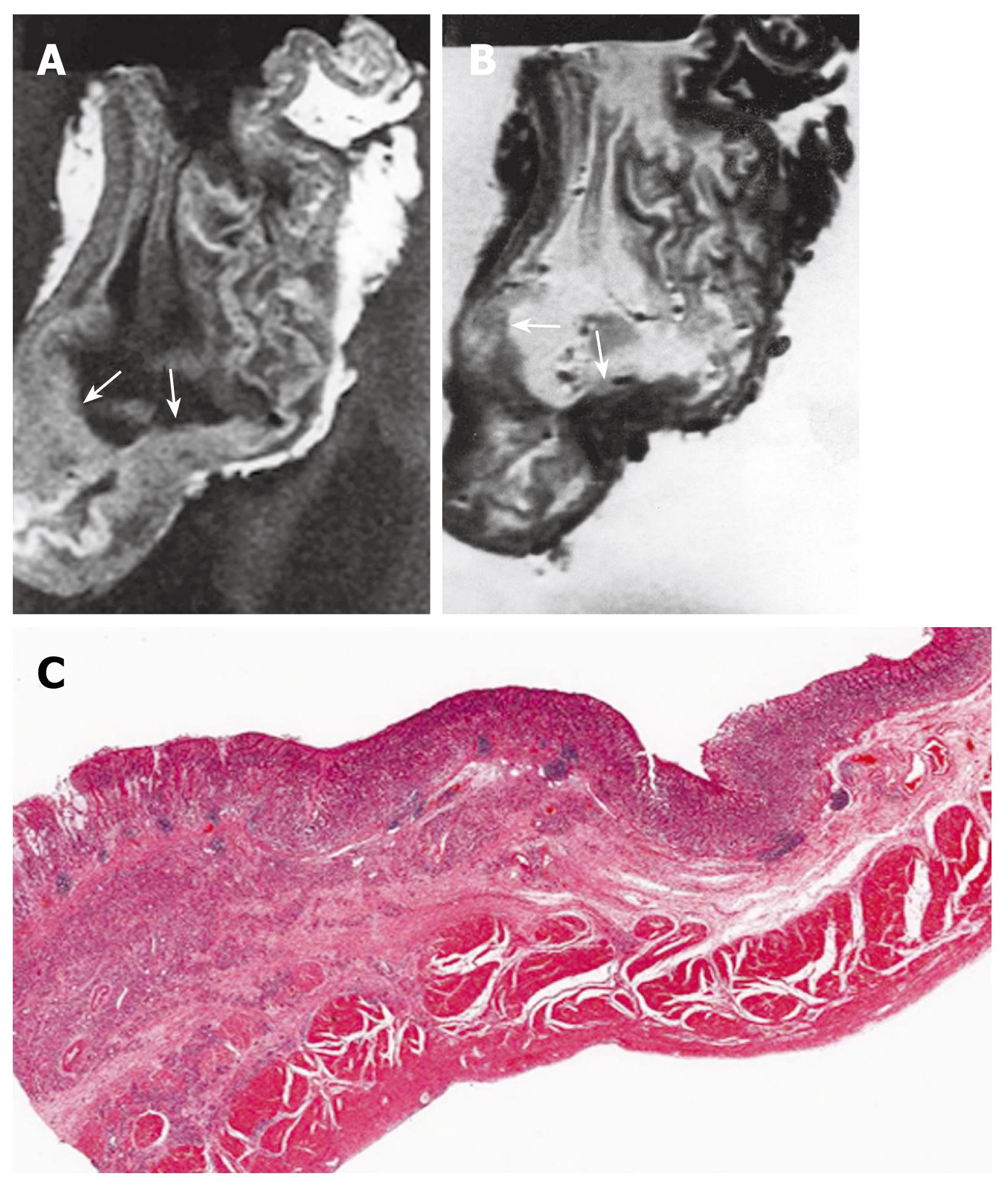
Figure 3 MRI and histology of T2 gastric cancer.
A: T1-weighted (500/20) sagittal image showed diffuse thickening of gastric wall with obliteration of mucosa, submucosa and muscularis propria SI in antrum and lower body, while preserved outer marginal SI (arrows); B: T2-weighted (2000/90) sagittal MRI showed ill defined lesion with minimal increased and same SI compared to surrounding normal gastric wall (arrows); C: Light microscopic section demonstrate proper muscle invasion of gastric cancer (HE stain; original magnification, × 1).
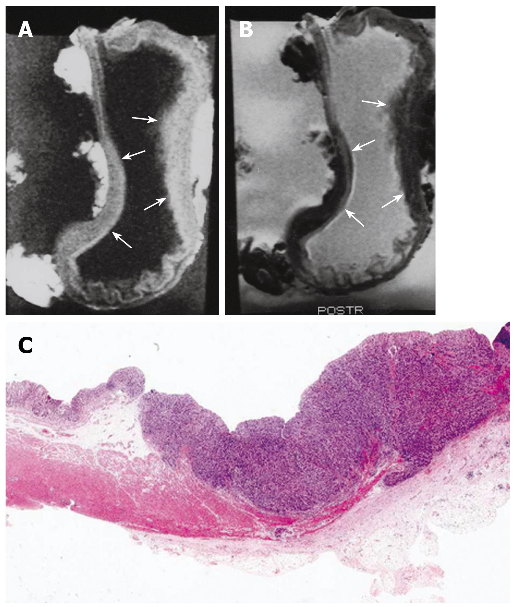
Figure 4 MRI and histology of T3 gastric adenocarcinoma.
A: T1-weighted (500/20) sagittal image showed thickening of gastric wall with all three layer SI change in lesser and greater curvature site of stomach body (arrows); B: T2-weighted (2000/90) sagittal MRI showed minimal increase of SI on lesion site and poor delineation of gastric wall SI at out layer margin compared to normal gastric wall (arrows); C: Light microscopic section showed extension of tumor invasion to serosal layer (HE stain; original magnification, × 1).
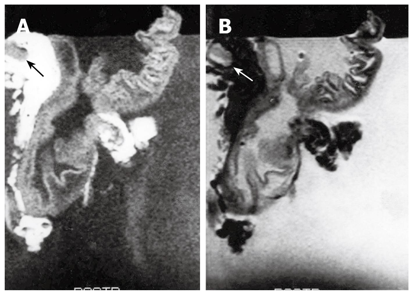
Figure 5 MRI of N1 gastric adenocarcinoma.
A: T1-weighted (500/20) MR image showed single lymph node on lesser curvature site of stomach body (arrow); B: T2-weighted (2000/90) MRI showed intermediated signal SI of lymph node (arrow).
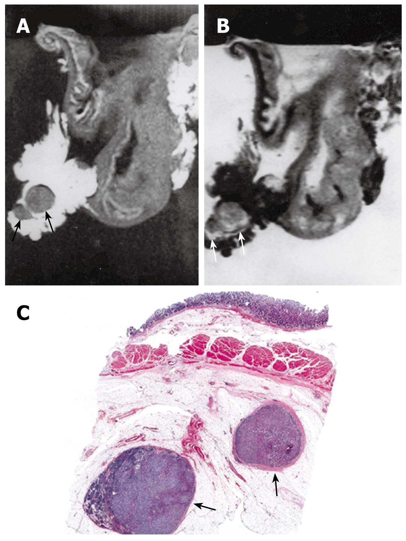
Figure 6 MRI and histology of N2 gastric adenocarcinoma.
A: T1-weighted (500/20) MRI showed two lymph nodes in lesser curvature site of stomach antrum (arrows). Eight lymph nodes are detected in total in perigastric area (not shown); B: T2-weighted (2000/90) MRI showed intermediate SI in the lymph nodes (arrows); C: Light microscopic section showed two lymph nodes in lesser curvature site of gastric antrum (arrows) (HE stain; original magnification, × 1).
- Citation: Kim IY, Kim SW, Shin HC, Lee MS, Jeong DJ, Kim CJ, Kim YT. MRI of gastric carcinoma: Results of T and N-staging in an in vitro study. World J Gastroenterol 2009; 15(32): 3992-3998
- URL: https://www.wjgnet.com/1007-9327/full/v15/i32/3992.htm
- DOI: https://dx.doi.org/10.3748/wjg.15.3992


