Copyright
©2009 The WJG Press and Baishideng.
World J Gastroenterol. Aug 21, 2009; 15(31): 3937-3939
Published online Aug 21, 2009. doi: 10.3748/wjg.15.3937
Published online Aug 21, 2009. doi: 10.3748/wjg.15.3937
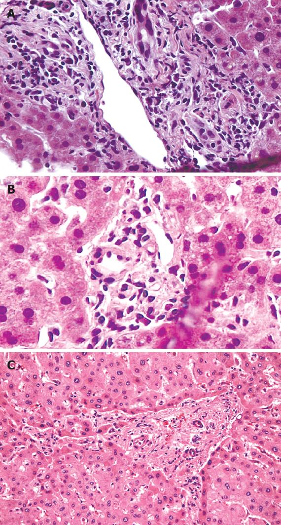
Figure 1 Hepatic histology.
A: Relatively large portal tract containing bile ducts with increased nuclear to cytoplasmic ratio, eosinophilic transformation of the cytoplasm, nuclear hyperchromasia, and uneven nuclear spacing; B: Small portal tract containing the hepatic artery and portal vein, but there is no bile duct. Five of twelve interlobular portal tracts in this biopsy lacked bile ducts; C: Hepatectomy specimen: loss of interlobular bile ducts in most of the small portal tracts, more advanced portal and periportal fibrosis with short fibrous septa. (Haematoxylin & Eosin stain, × 100).
- Citation: El Hajj II, Malik SM, Alwakeel HR, Shaikh OS, Sasatomi E, Kandil HM. Celecoxib-induced cholestatic liver failure requiring orthotopic liver transplantation. World J Gastroenterol 2009; 15(31): 3937-3939
- URL: https://www.wjgnet.com/1007-9327/full/v15/i31/3937.htm
- DOI: https://dx.doi.org/10.3748/wjg.15.3937


