Copyright
©2009 The WJG Press and Baishideng.
World J Gastroenterol. Aug 14, 2009; 15(30): 3831-3833
Published online Aug 14, 2009. doi: 10.3748/wjg.15.3831
Published online Aug 14, 2009. doi: 10.3748/wjg.15.3831
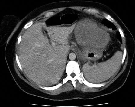
Figure 1 Computed tomography appearance of a well-circumscribed mass of mixed attenuation in the left upper abdomen, that was probably related to the stomach, and measured 10 cm in maximum diameter.
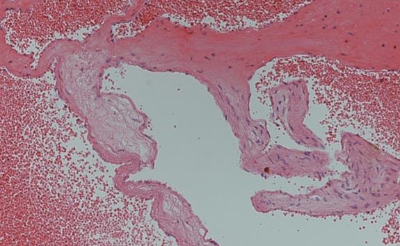
Figure 2 Numerous thick-walled blood vessels lined by endothelial cells and containing red blood cells.
Appearance was characteristic of cavernous hemangioma (HE, × 100).
- Citation: Chin KF, Khair G, Babu PS, Morgan DR. Cavernous hemangioma arising from the gastro-splenic ligament: A case report. World J Gastroenterol 2009; 15(30): 3831-3833
- URL: https://www.wjgnet.com/1007-9327/full/v15/i30/3831.htm
- DOI: https://dx.doi.org/10.3748/wjg.15.3831


