Copyright
©2009 The WJG Press and Baishideng.
World J Gastroenterol. Jan 21, 2009; 15(3): 378-379
Published online Jan 21, 2009. doi: 10.3748/wjg.15.378
Published online Jan 21, 2009. doi: 10.3748/wjg.15.378
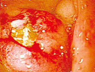
Figure 1 Gastroduodenal endoscopy revealed an ovoid mass in the first portion of the duodenum, the center of which harbored a mucosal defect.
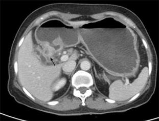
Figure 2 Abdominal CT showed wall thickening at the bulbar and postbulbar portions of the duodenum, with marked gastric dilatation.
A cystic lesion (arrow) was found at the thickened duodenal wall. The gallbladder was partially collapsed and adhered to the duodenum.
- Citation: Park SH, Lee SW, Song TJ. Another new variant of Bouveret’s syndrome. World J Gastroenterol 2009; 15(3): 378-379
- URL: https://www.wjgnet.com/1007-9327/full/v15/i3/378.htm
- DOI: https://dx.doi.org/10.3748/wjg.15.378


