Copyright
©2009 The WJG Press and Baishideng.
World J Gastroenterol. Aug 7, 2009; 15(29): 3704-3707
Published online Aug 7, 2009. doi: 10.3748/wjg.15.3704
Published online Aug 7, 2009. doi: 10.3748/wjg.15.3704
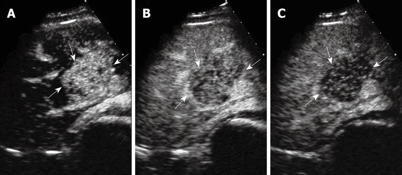
Figure 1 Real-time CEUS showing a solid, oval-shaped, hyperechogenic 5.
1 cm × 3.8 cm × 4.6 cm mass in the anterior segment of the right liver lobe. A: Hypervascularity in the arterial phase; B: Partial wash-out in the portal venous phase; C: Hypoechoic feature in the late phase.
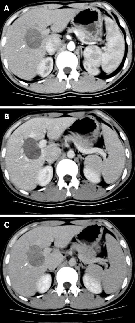
Figure 2 Transverse sequential triple-phase enhanced spiral CT showing a hypovascular mass in the anterior segment of the right liver lobe.
A: The arterial-dominant phase; B: The portal-dominant phase; C: The late phase.
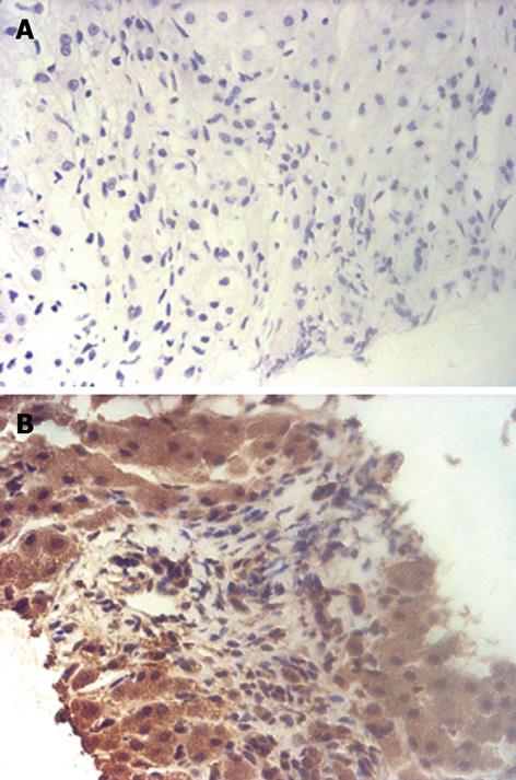
Figure 3 Histologic and immunohistochemical examinations of the tumor biopsy specimen.
A: Neoplastic spindle cells in an irregular pattern, with normal hepatic parenchyma in peripheral areas (HE, original magnification, × 200); B: Diffuse and strong positivity for CD117 (original magnification, × 200).
- Citation: Luo XL, Liu D, Yang JJ, Zheng MW, Zhang J, Zhou XD. Primary gastrointestinal stromal tumor of the liver: A case report. World J Gastroenterol 2009; 15(29): 3704-3707
- URL: https://www.wjgnet.com/1007-9327/full/v15/i29/3704.htm
- DOI: https://dx.doi.org/10.3748/wjg.15.3704


