Copyright
©2009 The WJG Press and Baishideng.
World J Gastroenterol. Aug 7, 2009; 15(29): 3697-3700
Published online Aug 7, 2009. doi: 10.3748/wjg.15.3697
Published online Aug 7, 2009. doi: 10.3748/wjg.15.3697
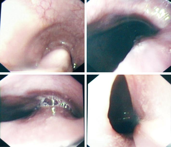
Figure 1 Endoscopy showing the long broad pedicle and broad base.
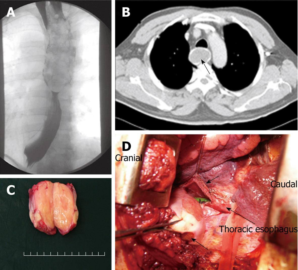
Figure 2 Recurrent Giant Esophageal polyp in 61-year-old man.
A: Barium swallow study; B: CT Thorax, showing the homogenous nature of the polyp (arrow); C: Bi-valved bulk of the polyp without its pedicle, showing the fatty nature of the polyp; D: Intra-operative trans-thoracic view showing the polyp’s head and an ulcer (bottom arrow), esophagotomy (top arrow) with the naso-gastric tube in-situ (middle arrow).
- Citation: Lee SY, Chan WH, Sivanandan R, Lim DTH, Wong WK. Recurrent giant fibrovascular polyp of the esophagus. World J Gastroenterol 2009; 15(29): 3697-3700
- URL: https://www.wjgnet.com/1007-9327/full/v15/i29/3697.htm
- DOI: https://dx.doi.org/10.3748/wjg.15.3697


