Copyright
©2009 The WJG Press and Baishideng.
World J Gastroenterol. Jul 21, 2009; 15(27): 3434-3436
Published online Jul 21, 2009. doi: 10.3748/wjg.15.3434
Published online Jul 21, 2009. doi: 10.3748/wjg.15.3434
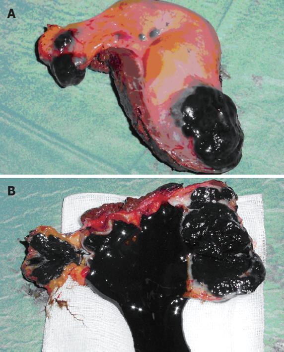
Figure 1 Surgical specimen.
A: Ulcerated black mass in the fundus and the cystic duct of the resected gallbladder; B: Gallbladder filled with black sludge and necrotic material.
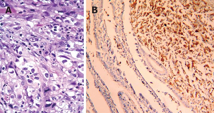
Figure 2 Pathological examination.
A: Microscopic picture of metastatic melanoma in the gallbladder (HE, × 20); B: Neoplastic cells immunostained with anti-S-100 antibodies (× 400).
- Citation: Vernadakis S, Rallis G, Danias N, Serafimidis C, Christodoulou E, Troullinakis M, Legakis N, Peros G. Metastatic melanoma of the gallbladder: An unusual clinical presentation of acute cholecystitis. World J Gastroenterol 2009; 15(27): 3434-3436
- URL: https://www.wjgnet.com/1007-9327/full/v15/i27/3434.htm
- DOI: https://dx.doi.org/10.3748/wjg.15.3434


