Copyright
©2009 The WJG Press and Baishideng.
World J Gastroenterol. Jul 21, 2009; 15(27): 3411-3416
Published online Jul 21, 2009. doi: 10.3748/wjg.15.3411
Published online Jul 21, 2009. doi: 10.3748/wjg.15.3411
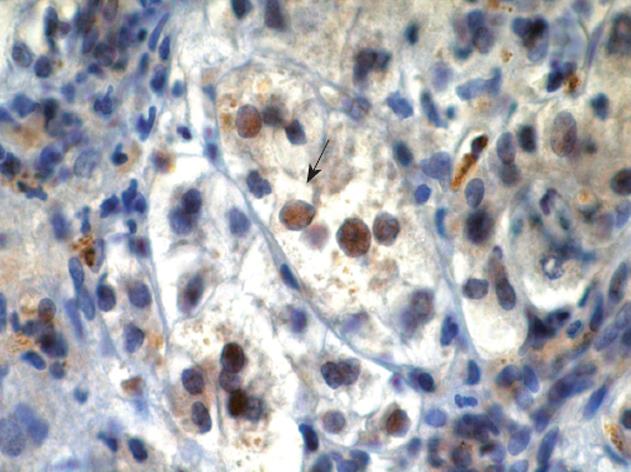
Figure 1 Immunohistochemistry of a positive case, consisting of brown colored nuclei (arrow), using a conventional optical microscope (× 640).
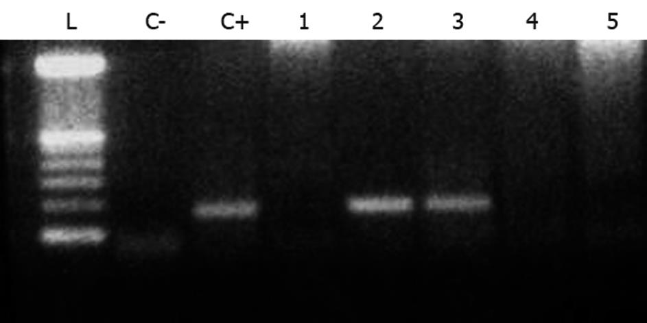
Figure 2 HCMV DNA results of 2% agarose gel electrophoresis under ultraviolet light (PCR).
L: Ladder; C+: Positive control; C-: Negative control; 1, 4 and 5: Negative samples; 2 and 3: Positive samples.
- Citation: Bellomo-Brandao MA, Andrade PD, Costa SC, Escanhoela CA, Vassallo J, Porta G, De Tommaso AM, Hessel G. Cytomegalovirus frequency in neonatal intrahepatic cholestasis determined by serology, histology, immunohistochemistry and PCR. World J Gastroenterol 2009; 15(27): 3411-3416
- URL: https://www.wjgnet.com/1007-9327/full/v15/i27/3411.htm
- DOI: https://dx.doi.org/10.3748/wjg.15.3411


