Copyright
©2009 The WJG Press and Baishideng.
World J Gastroenterol. Jul 7, 2009; 15(25): 3178-3182
Published online Jul 7, 2009. doi: 10.3748/wjg.15.3178
Published online Jul 7, 2009. doi: 10.3748/wjg.15.3178
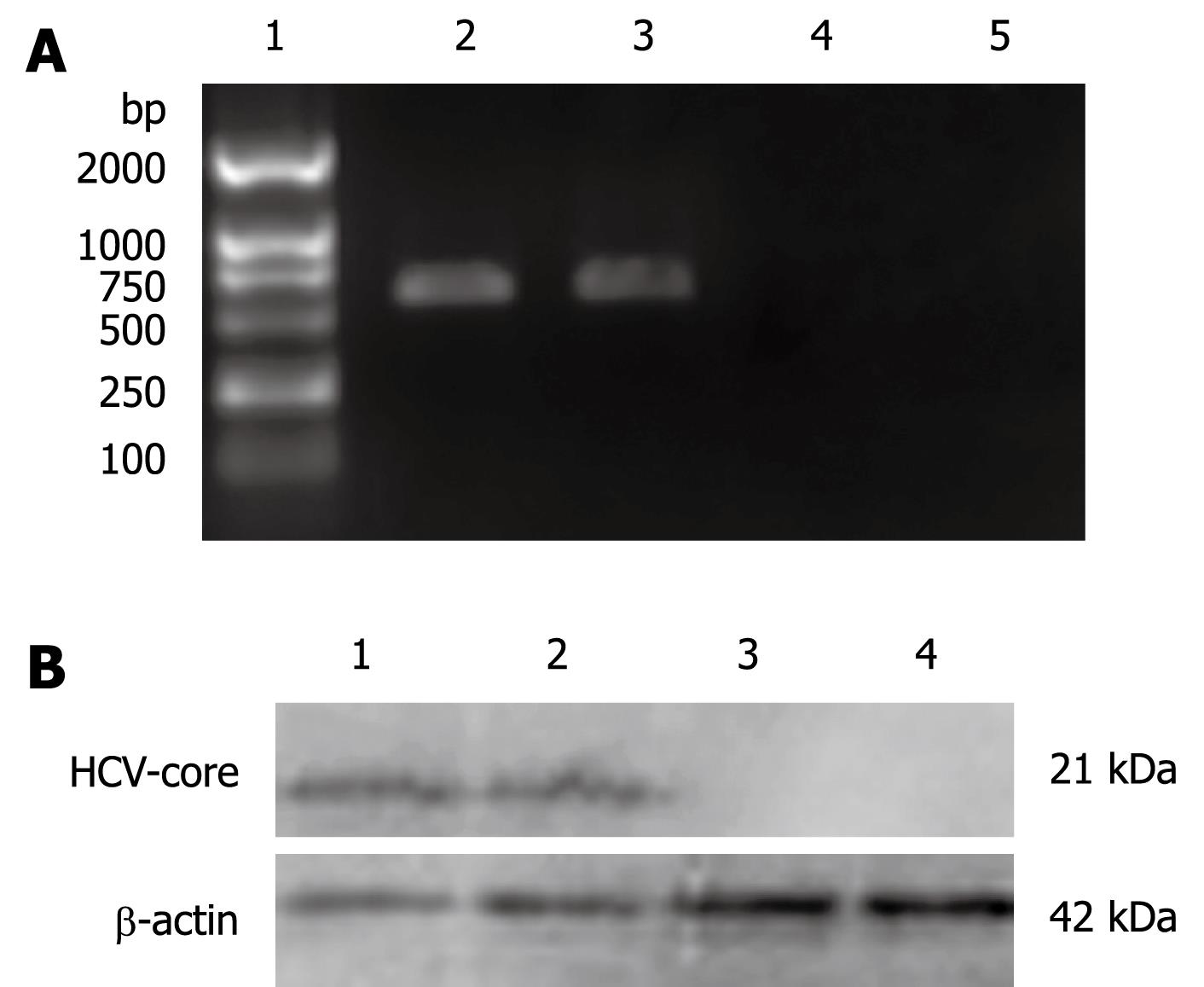
Figure 1 Expression of HCV-core in the stable cell line L02/core.
A: Detection of HCV-core mRNA by RT-PCR. 572 bp visible fragments consistent with the predicted size of HCV-core mRNA were detected in L02/core cell clones. 1: DL2000 marker; 2, 3: L02 cells transfected with pcDNA3.1-core (L02/core); 4: L02 cells transfected with pcDNA3.1 (empty vector); 5: Untransfected L02 cells; B: Detection of HCV-core protein by Western blotting. 1, 2: L02/core cells; 3: L02 cells transfected with empty vector; 4: Untransfected L02 cells.
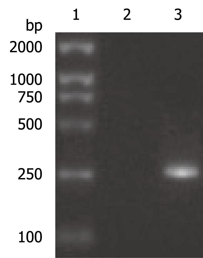
Figure 2 PCR product of OAS promoter.
A 240 bp visible fragment consistent with the predicted size was observed by electrophoresis. 1: DL2000 marker; 2: Negative control; 3: PCR product.
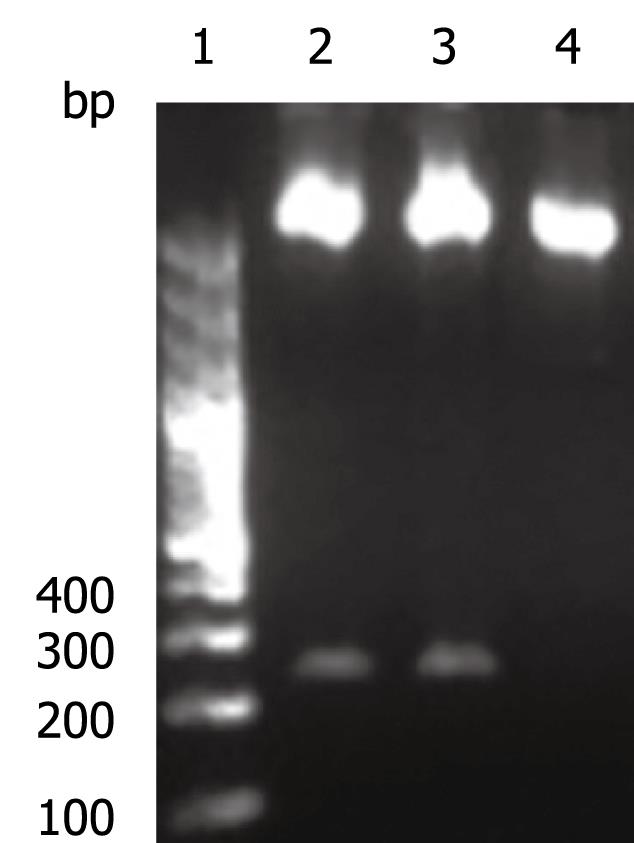
Figure 3 The OAS-luciferase construct (pGL3-OAS-Luci) was confirmed by double enzyme digestion with SacI and HindIII.
1: GeneRuler™ 100 bp DNA Ladder; 2-3: Double digestion of pGL3-OAS-Luci; 4: Double digestion of pGL3-Basic.
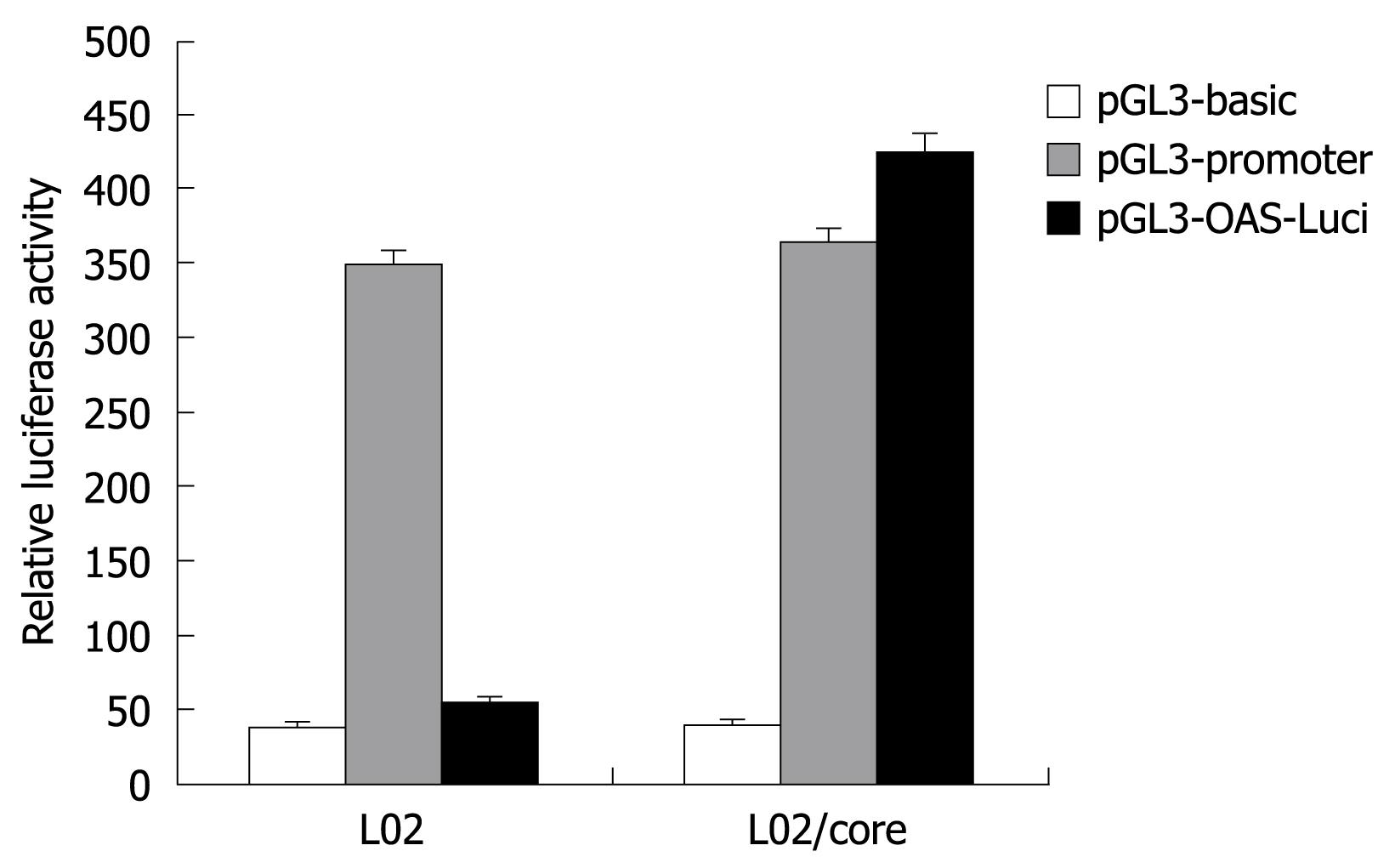
Figure 4 Transcriptional activity of the OAS promoter in L02 and L02/core cells.
The OAS promoter showed significant transcriptional activity in the presence of HCV-core protein (L02/core), but not in the normal liver cells (L02). The data shown are the mean ± SD from three independent experiments.
- Citation: Wang Y, Mao SS, He QQ, Zi Y, Wen JF, Feng DY. Specific activation of 2'-5'oligoadenylate synthetase gene promoter by hepatitis C virus-core protein: A potential for developing hepatitis C virus targeting gene therapy. World J Gastroenterol 2009; 15(25): 3178-3182
- URL: https://www.wjgnet.com/1007-9327/full/v15/i25/3178.htm
- DOI: https://dx.doi.org/10.3748/wjg.15.3178


