Copyright
©2009 The WJG Press and Baishideng.
World J Gastroenterol. Jun 28, 2009; 15(24): 3003-3008
Published online Jun 28, 2009. doi: 10.3748/wjg.15.3003
Published online Jun 28, 2009. doi: 10.3748/wjg.15.3003
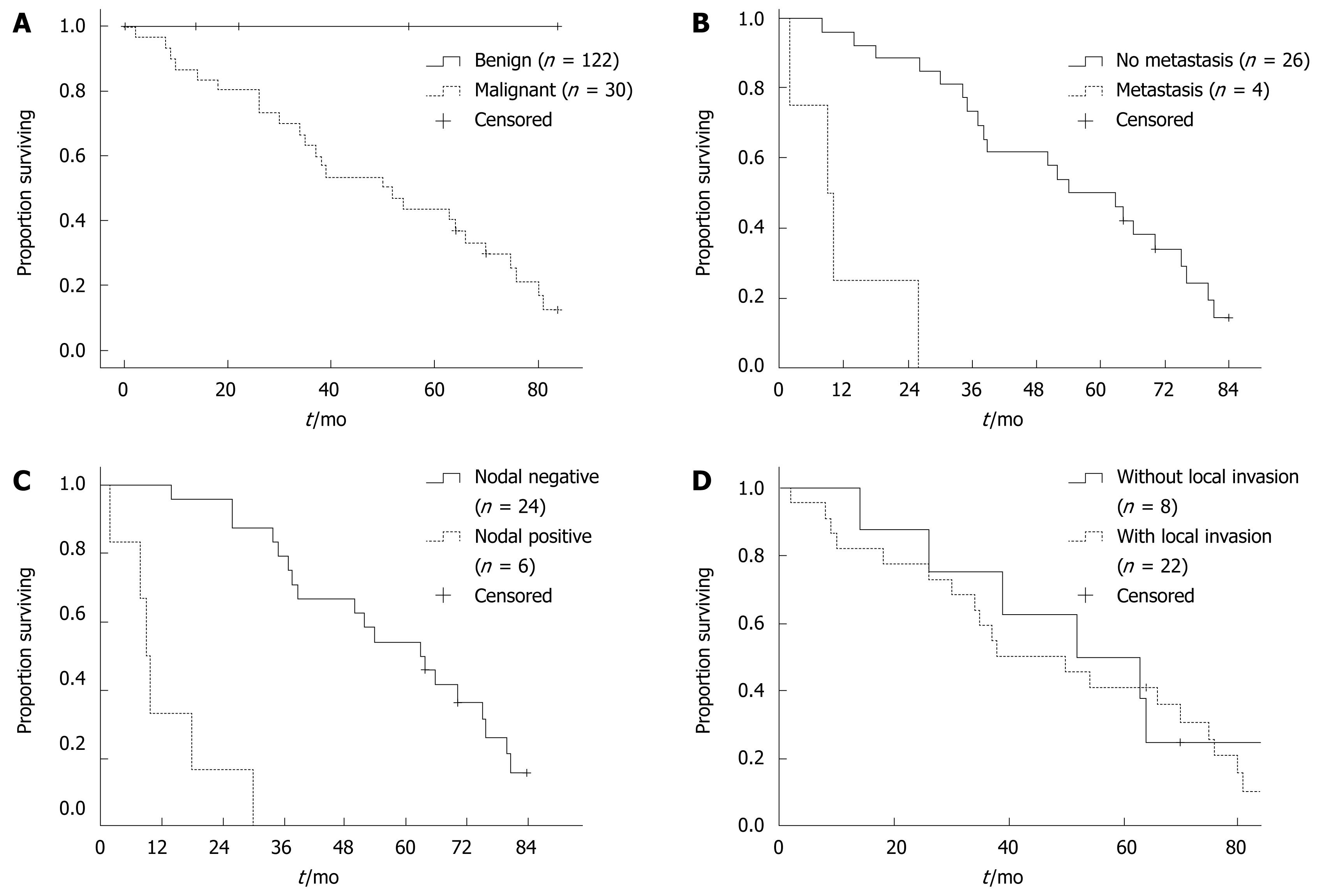
Figure 1 Survival of patients undergoing resection of paraganglioma.
Stratified by A: Malignancy (P < 0.001); Stratified by B: Metastasis (P < 0.001); Stratified by C: Nodal status (P < 0.001); Stratified by D: Local invasion (P = 0.708).
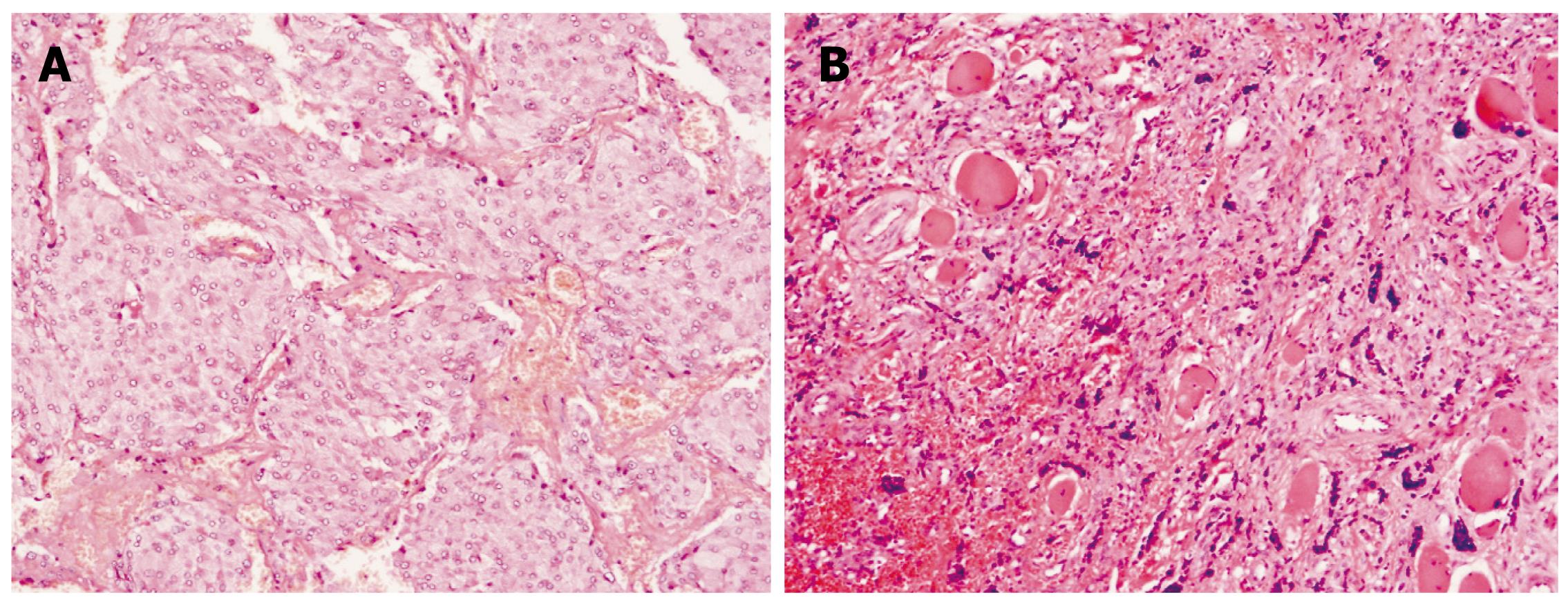
Figure 2 The histologic analysis of paraganglioma.
The hematoxylin-eosin stain of chief (type 1) cells arranged in one of the typical cell nests in a retroperitoneal benign tumor (A), and in a femur metastasis (B), (× 50).
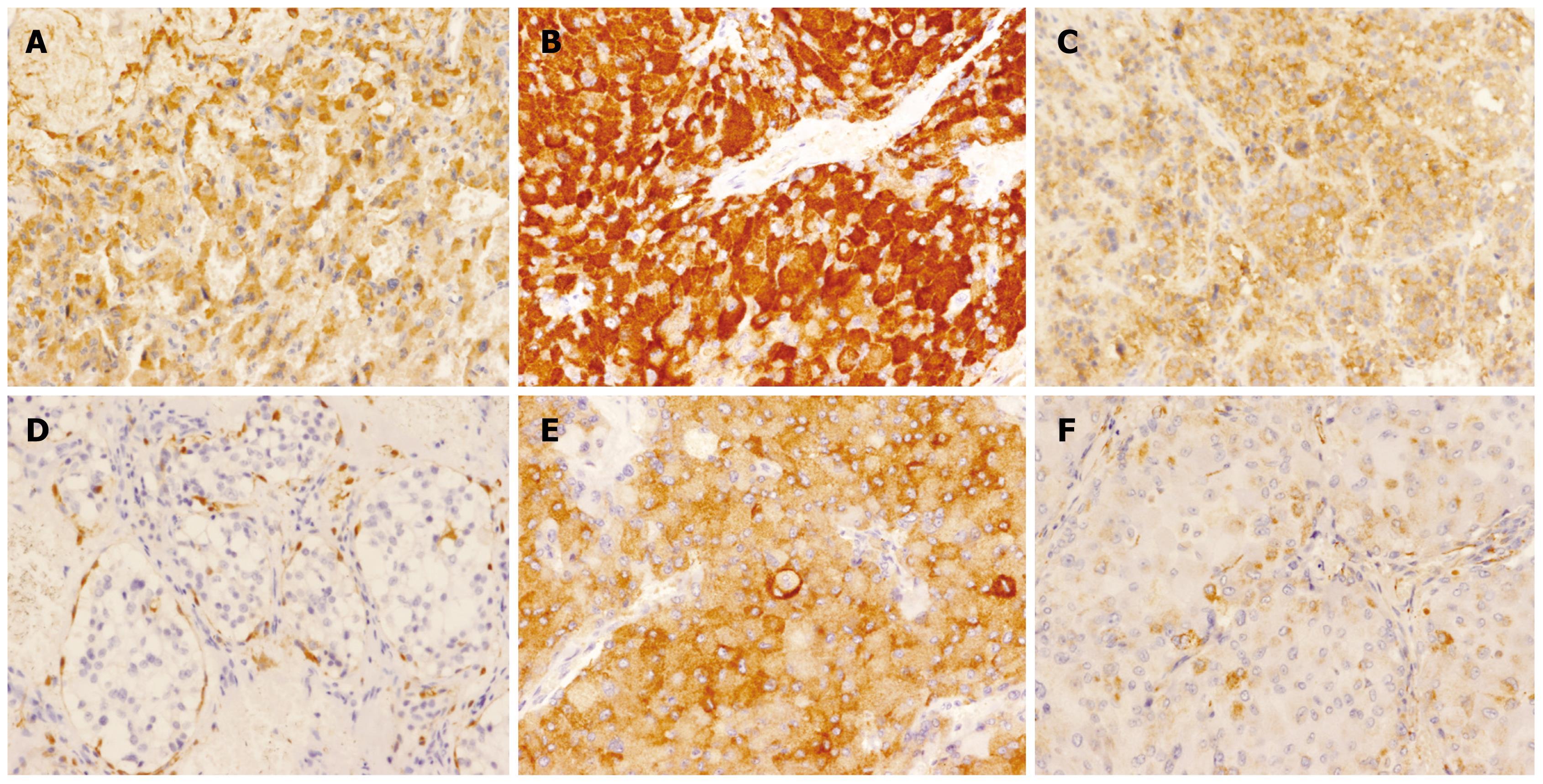
Figure 3 The immunohistochemistry analysis of paraganglioma.
The immunolabeling test shows that peripheral to the cell nests are the stellate sustentacular (type 2) cells, intensely brown after S-100 immunolabeling (D), and in some cases CK (A, CgA (B), NSE (C), Syn (E) and vimentin (F) are also positive for paraganglioma, (× 100).
- Citation: Feng N, Zhang WY, Wu XT. Clinicopathological analysis of paraganglioma with literature review. World J Gastroenterol 2009; 15(24): 3003-3008
- URL: https://www.wjgnet.com/1007-9327/full/v15/i24/3003.htm
- DOI: https://dx.doi.org/10.3748/wjg.15.3003


